 Movie
Movie Controller

[English] 日本語
 Yorodumi
- PDB-2wnx: 3b' carbohydrate-binding module from the Cel9V glycoside hydrolas... -
+ Open data

- Basic information
| Entry | Database: PDB / ID: 2wnx | ||||||
|---|---|---|---|---|---|---|---|
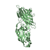
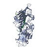
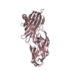
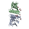
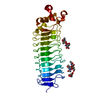
| Title | 3b' carbohydrate-binding module from the Cel9V glycoside hydrolase from Clostridium thermocellum | ||||||
 Components Components | GLYCOSIDE HYDROLASE, FAMILY 9 | ||||||
 Keywords Keywords | HYDROLASE / CELLULOSE DEGRADATION / GLYCOSIDE HYDROLASE | ||||||
| Function / homology |  Function and homology information Function and homology informationcellulose binding / polysaccharide catabolic process / hydrolase activity, hydrolyzing O-glycosyl compounds / metal ion binding Similarity search - Function | ||||||
| Biological species |  CLOSTRIDIUM THERMOCELLUM (bacteria) CLOSTRIDIUM THERMOCELLUM (bacteria) | ||||||
| Method |  X-RAY DIFFRACTION / SYNCHROTRON / MOLECULAR REPLACEMENT / Resolution: 1.31 Å X-RAY DIFFRACTION / SYNCHROTRON / MOLECULAR REPLACEMENT / Resolution: 1.31 Å | ||||||
 Authors Authors | Petkun, S. / Jindou, S. / Shimon, L.J.W. / Bayer, E.A. / Lamed, R. / Frolow, F. | ||||||
 Citation Citation | Journal: Acta Crystallogr.,Sect.D / Year: 2010 Title: Structure of a Family 3B' Carbohydrate-Binding Module from the Cel9V Glycoside Hydrolase from Clostridium Thermocellum: Structural Diversity and Implications for Carbohydrate Binding Authors: Petkun, S. / Jindou, S. / Shimon, L.J.W. / Bayer, E.A. / Lamed, R. / Frolow, F. #1: Journal: Fems Microbiol.Lett. / Year: 2006 Title: Novel Architecture of Family-9 Glycoside Hydrolases Identified in Cellulosomal Enzymes of Acetivibrio Cellulolyticus and Clostridium Thermocellum. Authors: Jindou, S. / Xu, Q. / Kenig, R. / Shulman, M. / Shoham, Y. / Bayer, E.A. / Lamed, R. | ||||||
| History |
|
- Structure visualization
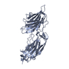
| Structure viewer | Molecule: MolmilJmol/JSmol |
|---|
- Downloads & links
-Download
| PDBx/mmCIF format | 2wnx.cif.gz | 96.4 KB | Display | PDBx/mmCIF format |
|---|---|---|---|---|
| PDB format | pdb2wnx.ent.gz | 72.4 KB | Display | PDB format |
| PDBx/mmJSON format | 2wnx.json.gz | Tree view | PDBx/mmJSON format | |
| Others |  Other downloads Other downloads |
-Validation report
| Arichive directory | https://data.pdbj.org/pub/pdb/validation_reports/wn/2wnxftp://data.pdbj.org/pub/pdb/validation_reports/wn/2wnx | HTTPS FTP |
|---|
-Related structure data
| Related structure data |  2wo4C  2wobC  1nbcS C: citing same article ( S: Starting model for refinement |
|---|---|
| Similar structure data |


-Links
 PDBj
PDBj

- Assembly
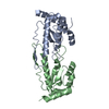
| Deposited unit | 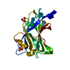
| ||||||||
|---|---|---|---|---|---|---|---|---|---|
| 1 | 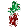
| ||||||||
| Unit cell |
|
-Components
| #1: Protein | Mass: 18649.406 Da / Num. of mol.: 1 / Fragment: CARBOHYDRATE-BINDING MODULE3B', RESIDUES 731-888 Source method: isolated from a genetically manipulated source Source: (gene. exp.) CLOSTRIDIUM THERMOCELLUM (bacteria) / Production host: | ||||
|---|---|---|---|---|---|
| #2: Chemical | ChemComp-CA /   Mass: 40.078 Da / Num. of mol.: 1 / Source method: obtained synthetically / Formula: Ca Mass: 40.078 Da / Num. of mol.: 1 / Source method: obtained synthetically / Formula: Ca | ||||
| #3: Chemical |   Mass: 96.063 Da / Num. of mol.: 3 / Source method: obtained synthetically / Formula: SO4 Mass: 96.063 Da / Num. of mol.: 3 / Source method: obtained synthetically / Formula: SO4#4: Chemical | ChemComp-FMT / |   Mass: 46.025 Da / Num. of mol.: 1 / Source method: obtained synthetically / Formula: CH2O2 Mass: 46.025 Da / Num. of mol.: 1 / Source method: obtained synthetically / Formula: CH2O2#5: Water | ChemComp-HOH / |  Mass: 18.015 Da / Num. of mol.: 349 / Source method: isolated from a natural source / Formula: H2O Mass: 18.015 Da / Num. of mol.: 349 / Source method: isolated from a natural source / Formula: H2O |
-Experimental details
-Experiment
| Experiment | Method: X-RAY DIFFRACTION / Number of used crystals: 1 |
|---|
- Sample preparation
| Crystal | Density Matthews: 3.52 Å3/Da / Density % sol: 65 % / Description: MONOMER A |
|---|---|
| Crystal grow | pH: 5.6 Details: 0.2 M AMMONIUM ACETATE, 0.1 M TRI-SODIUM CITRATE DIHYDRATE PH 5.6, 30% W/V POLYETHYLENE GLYCOL 4000 |
-Data collection
| Diffraction | Mean temperature: 100 K |
|---|---|
| Diffraction source | Source: SYNCHROTRON / Site: ESRF  / Beamline: ID29 / Wavelength: 0.9756 / Beamline: ID29 / Wavelength: 0.9756 |
| Detector | Type: ADSC CCD / Detector: CCD / Date: Dec 19, 2004 |
| Radiation | Protocol: SINGLE WAVELENGTH / Monochromatic (M) / Laue (L): M / Scattering type: x-ray |
| Radiation wavelength | Wavelength: 0.9756 Å / Relative weight: 1 |
| Reflection | Resolution: 1.3→50 Å / Num. obs: 57111 / % possible obs: 94.6 % / Observed criterion σ(I): 0 / Redundancy: 8.1 % / Biso Wilson estimate: 17.1 Å2 / Rmerge(I) obs: 0.07 / Net I/σ(I): 48.1 |
| Reflection shell | Resolution: 1.3→1.35 Å / Redundancy: 7.6 % / Rmerge(I) obs: 0.52 / Mean I/σ(I) obs: 1.86 / % possible all: 81.5 |
- Processing
| Software |
| ||||||||||||||||||||||||||||||||||||||||||||||||||||||||||||||||||||||||||||||||||||||||||||||||||||||||||||||||||||||||||||||||||||||||||||||||||||||||||||||||||||||||||||||||||||||
|---|---|---|---|---|---|---|---|---|---|---|---|---|---|---|---|---|---|---|---|---|---|---|---|---|---|---|---|---|---|---|---|---|---|---|---|---|---|---|---|---|---|---|---|---|---|---|---|---|---|---|---|---|---|---|---|---|---|---|---|---|---|---|---|---|---|---|---|---|---|---|---|---|---|---|---|---|---|---|---|---|---|---|---|---|---|---|---|---|---|---|---|---|---|---|---|---|---|---|---|---|---|---|---|---|---|---|---|---|---|---|---|---|---|---|---|---|---|---|---|---|---|---|---|---|---|---|---|---|---|---|---|---|---|---|---|---|---|---|---|---|---|---|---|---|---|---|---|---|---|---|---|---|---|---|---|---|---|---|---|---|---|---|---|---|---|---|---|---|---|---|---|---|---|---|---|---|---|---|---|---|---|---|---|
| Refinement | Method to determine structure: MOLECULAR REPLACEMENT Starting model: PDB ENTRY 1NBC Resolution: 1.31→20 Å / Cor.coef. Fo:Fc: 0.981 / Cor.coef. Fo:Fc free: 0.963 / SU B: 1.011 / SU ML: 0.019 / Cross valid method: THROUGHOUT / ESU R: 0.034 / ESU R Free: 0.037 / Stereochemistry target values: MAXIMUM LIKELIHOOD Details: HYDROGENS HAVE BEEN ADDED IN THE RIDING POSITIONS. ATOM RECORD CONTAINS SUM OF TLS AND RESIDUAL B FACTORS U VALUES WITH TLS ADDED DISORDERED REGIONS WERE MODELED STEREOCHEMICALLY
| ||||||||||||||||||||||||||||||||||||||||||||||||||||||||||||||||||||||||||||||||||||||||||||||||||||||||||||||||||||||||||||||||||||||||||||||||||||||||||||||||||||||||||||||||||||||
| Solvent computation | Ion probe radii: 0.8 Å / Shrinkage radii: 0.8 Å / VDW probe radii: 1.4 Å / Solvent model: BABINET MODEL WITH MASK | ||||||||||||||||||||||||||||||||||||||||||||||||||||||||||||||||||||||||||||||||||||||||||||||||||||||||||||||||||||||||||||||||||||||||||||||||||||||||||||||||||||||||||||||||||||||
| Displacement parameters | Biso mean: 16.301 Å2
| ||||||||||||||||||||||||||||||||||||||||||||||||||||||||||||||||||||||||||||||||||||||||||||||||||||||||||||||||||||||||||||||||||||||||||||||||||||||||||||||||||||||||||||||||||||||
| Refinement step | Cycle: LAST / Resolution: 1.31→20 Å
| ||||||||||||||||||||||||||||||||||||||||||||||||||||||||||||||||||||||||||||||||||||||||||||||||||||||||||||||||||||||||||||||||||||||||||||||||||||||||||||||||||||||||||||||||||||||
| Refine LS restraints |
|