 Movie
Movie Controller

[English] 日本語
 Yorodumi
- PDB-2wmp: Structure of the E. coli chaperone PapD in complex with the pilin... -
+ Open data

- Basic information
| Entry | Database: PDB / ID: 2wmp | ||||||
|---|---|---|---|---|---|---|---|
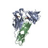
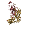
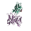
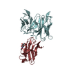
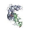
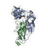
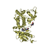
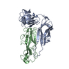
| Title | Structure of the E. coli chaperone PapD in complex with the pilin domain of the PapGII adhesin | ||||||
 Components Components |
| ||||||
 Keywords Keywords | CHAPERONE / CELL ADHESION / DONOR STRAND COMPLEMENTATION / PILIN DOMAIN / IMMUNOGLOBULIN DOMAIN / BACTERIAL ATTACHMENT AND INVASION / DONOR STRAND EXCHANGE / CHAPERONE USHER PATHWAY | ||||||
| Function / homology |  Function and homology information Function and homology informationpilus / cell wall organization / outer membrane-bounded periplasmic space / carbohydrate binding / cell adhesion Similarity search - Function | ||||||
| Biological species |  | ||||||
| Method |  X-RAY DIFFRACTION / SYNCHROTRON / MOLECULAR REPLACEMENT / Resolution: 2.3 Å X-RAY DIFFRACTION / SYNCHROTRON / MOLECULAR REPLACEMENT / Resolution: 2.3 Å | ||||||
 Authors Authors | Ford, B.A. / Verger, D. / Elam, J.S. / Dodson, K.W. / Pinkner, J.S. / Hultgren, S.J. | ||||||
 Citation Citation | Journal: J.Bacteriol. / Year: 2012 Title: Structure of the Papd-Papgii Pilin Complex Reveals an Open and Flexible P5 Pocket. Authors: Ford, B.A. / Verger, D. / Dodson, K.W. / Volkan, E. / Kostakioti, M. / Elam, J.S. / Pinkner, J.S. / Waksman, G. / Hultgren, S.J. #1: Journal: Cell(Cambridge,Mass.) / Year: 2001Title: Structural Basis of the Interaction of the Pyelonephritic E. Coli Adhesin to its Human Kidney Receptor Authors: Dodson, K.W. / Pinkner, J.S. / Rose, T. / Magnusson, G. / Hultgren, S.J. / Waksman, G. | ||||||
| History |
| ||||||
| Remark 700 | SHEET THE SHEET STRUCTURE OF THIS MOLECULE IS BIFURCATED. IN ORDER TO REPRESENT THIS FEATURE IN ... SHEET THE SHEET STRUCTURE OF THIS MOLECULE IS BIFURCATED. IN ORDER TO REPRESENT THIS FEATURE IN THE SHEET RECORDS BELOW, TWO SHEETS ARE DEFINED. |
- Structure visualization
| Structure viewer | Molecule: MolmilJmol/JSmol |
|---|
- Downloads & links
-Download
| PDBx/mmCIF format | 2wmp.cif.gz | 81.5 KB | Display | PDBx/mmCIF format |
|---|---|---|---|---|
| PDB format | pdb2wmp.ent.gz | 60.2 KB | Display | PDB format |
| PDBx/mmJSON format | 2wmp.json.gz | Tree view | PDBx/mmJSON format | |
| Others |  Other downloads Other downloads |
-Validation report
| Arichive directory | https://data.pdbj.org/pub/pdb/validation_reports/wm/2wmpftp://data.pdbj.org/pub/pdb/validation_reports/wm/2wmp | HTTPS FTP |
|---|
-Related structure data
| Related structure data |  1pdkS S: Starting model for refinement |
|---|---|
| Similar structure data |


-Links
 PDBj
PDBj
- Assembly
| Deposited unit | 
| ||||||||
|---|---|---|---|---|---|---|---|---|---|
| 1 |
| ||||||||
| Unit cell |
|
-Components
| #1: Protein | Mass: 25418.777 Da / Num. of mol.: 1 / Fragment: RESIDUES 22-239 Source method: isolated from a genetically manipulated source Details: C TERMINUS 6-HISTIDINE TAG / Source: (gene. exp.) |
|---|---|
| #2: Protein | Mass: 13136.854 Da / Num. of mol.: 1 / Fragment: PILIN DOMAIN, RESIDUES 214-336 Source method: isolated from a genetically manipulated source Details: ADHESIN DOMAIN DELETED (RESIDUES 1 TO 193) / Source: (gene. exp.) |
| #3: Water | ChemComp-HOH /  Mass: 18.015 Da / Num. of mol.: 192 / Source method: isolated from a natural source / Formula: H2O Mass: 18.015 Da / Num. of mol.: 192 / Source method: isolated from a natural source / Formula: H2O |
| Has protein modification | Y |
-Experimental details
-Experiment
| Experiment | Method: X-RAY DIFFRACTION / Number of used crystals: 1 |
|---|
- Sample preparation
| Crystal | Density Matthews: 2.52 Å3/Da / Density % sol: 51 % / Description: NONE |
|---|---|
| Crystal grow | pH: 8.5 Details: PROTEIN (COMPLEX) AT 9.9 MG/ML 10-12% PEG 4K 0.1M TRIS-HCL PH 8.5 5% ISOPROPANOL |
-Data collection
| Diffraction | Mean temperature: 100 K |
|---|---|
| Diffraction source | Source: SYNCHROTRON / Site: ESRF  / Beamline: ID23-2 / Wavelength: 0.8726 / Beamline: ID23-2 / Wavelength: 0.8726 |
| Detector | Type: ADSC CCD / Detector: CCD / Date: Apr 24, 2008 |
| Radiation | Protocol: SINGLE WAVELENGTH / Monochromatic (M) / Laue (L): M / Scattering type: x-ray |
| Radiation wavelength | Wavelength: 0.8726 Å / Relative weight: 1 |
| Reflection | Resolution: 2.3→66.56 Å / Num. obs: 17935 / % possible obs: 100 % / Observed criterion σ(I): 1 / Redundancy: 6.6 % / Biso Wilson estimate: 32.2 Å2 / Rmerge(I) obs: 0.13 / Net I/σ(I): 4.6 |
| Reflection shell | Resolution: 2.3→2.42 Å / Redundancy: 6.7 % / Rmerge(I) obs: 0.5 / Mean I/σ(I) obs: 1.4 / % possible all: 100 |
- Processing
| Software |
| ||||||||||||||||||||||||||||||||||||||||||||||||||||||||||||||||||||||||||||||||
|---|---|---|---|---|---|---|---|---|---|---|---|---|---|---|---|---|---|---|---|---|---|---|---|---|---|---|---|---|---|---|---|---|---|---|---|---|---|---|---|---|---|---|---|---|---|---|---|---|---|---|---|---|---|---|---|---|---|---|---|---|---|---|---|---|---|---|---|---|---|---|---|---|---|---|---|---|---|---|---|---|---|
| Refinement | Method to determine structure: MOLECULAR REPLACEMENT Starting model: PDB ENTRY 1PDK Resolution: 2.3→200 Å / Rfactor Rfree error: 0.01 / Data cutoff high absF: 10000 / Data cutoff low absF: 0 / Isotropic thermal model: RESTRAINED / Cross valid method: THROUGHOUT / σ(F): 0 / Stereochemistry target values: MAXIMUM LIKELIHOOD Details: THIS PDB FILE IS LINKED TO ENTRY 3ME0 AS BOTH FILES WILL APPEAR IN THE SAME PUBLICATION.
| ||||||||||||||||||||||||||||||||||||||||||||||||||||||||||||||||||||||||||||||||
| Solvent computation | Solvent model: DENSITY MODIFICATION / Bsol: 25.1473 Å2 / ksol: 0.3 e/Å3 | ||||||||||||||||||||||||||||||||||||||||||||||||||||||||||||||||||||||||||||||||
| Displacement parameters |
| ||||||||||||||||||||||||||||||||||||||||||||||||||||||||||||||||||||||||||||||||
| Refinement step | Cycle: LAST / Resolution: 2.3→200 Å
| ||||||||||||||||||||||||||||||||||||||||||||||||||||||||||||||||||||||||||||||||
| Refine LS restraints |
| ||||||||||||||||||||||||||||||||||||||||||||||||||||||||||||||||||||||||||||||||
| LS refinement shell | Resolution: 2.3→2.34 Å / Rfactor Rfree error: 0.054 / Total num. of bins used: 18
| ||||||||||||||||||||||||||||||||||||||||||||||||||||||||||||||||||||||||||||||||
| Xplor file |
|
