Mass: 18.015 Da / Num. of mol.: 570 / Source method: isolated from a natural source / Formula: H2O
Sequence details
PBP2B STRAIN R6 P0A3M6 PROTEIN SEQUENCE CODE IS THE CLOSER ONE TO MUTANT STRAIN 5204 (58 MUTATIONS). ...PBP2B STRAIN R6 P0A3M6 PROTEIN SEQUENCE CODE IS THE CLOSER ONE TO MUTANT STRAIN 5204 (58 MUTATIONS). THE PROTEIN SEQUENCE OF PBP2B STRAIN 5204 WAS OBTAINED BY OUR COLLABORATOR (REFERENCE 1 PAGLIERO ET AL. 2004, WITH OUT BEEN SUBMITED TO THE DATABASES). HOWEVER THE PBP2B 5204 TRANSPEPTIDASE DOMAIN ALONE AS BEEN SEQUENCE AND SUBMITED TO THE DATABASE (Q5ZGA5). LAST MODIFIED JULY 22, 2008. VERSION 14.
-
Experimental details
-
Experiment
Experiment
Method: X-RAY DIFFRACTION / Number of used crystals: 1
-
Sample preparation
Crystal
Density Matthews: 2.3 Å3/Da / Density % sol: 55 % / Description: NONE
Crystal grow
pH: 8 Details: 100 MM BIS-TRIS-HCL PH 5.5, 300 MM NACL, 5 MM ZN ACETATE, 30% W/V POLYETHYLENE GLYCOL MME 550
-
Data collection
Diffraction
ID
Mean temperature (K)
Crystal-ID
1
100
1
2
1
Diffraction source
Source
Site
Beamline
ID
Wavelength
Wavelength (Å)
SYNCHROTRON
ESRF
ID14-3
1
0.931
SYNCHROTRON
ESRF
BM30A
2
1.282740, 1.283334, 1.279625
Detector
Type
ID
Detector
Date
ADSC CCD
1
CCD
Jun 10, 2007
ADSC CCD
2
CCD
Radiation
ID
Protocol
Monochromatic (M) / Laue (L)
Scattering type
Wavelength-ID
1
SINGLEWAVELENGTH
M
x-ray
1
2
M
x-ray
1
Radiation wavelength
ID
Wavelength (Å)
Relative weight
1
0.931
1
2
1.28274
1
3
1.283334
1
4
1.279625
1
Reflection
Resolution: 2.18→44.5 Å / Num. obs: 108466 / % possible obs: 91 % / Observed criterion σ(I): 3 / Redundancy: 4.4 % / Biso Wilson estimate: 46.42 Å2 / Rsym value: 0.07 / Net I/σ(I): 17.34
Reflection shell
Resolution: 2.18→2.31 Å / Redundancy: 4 % / Mean I/σ(I) obs: 2.97 / Rsym value: 0.6 / % possible all: 70.1
-
Processing
Software
Name
Version
Classification
REFMAC
5.5.0070
refinement
XDS
datareduction
XSCALE
datascaling
autoSHARP
phasing
Refinement
Method to determine structure: MAD Starting model: NONE Resolution: 2.18→44.5 Å / Cor.coef. Fo:Fc: 0.946 / Cor.coef. Fo:Fc free: 0.916 / SU B: 15.858 / SU ML: 0.178 / TLS residual ADP flag: LIKELY RESIDUAL / Cross valid method: THROUGHOUT / ESU R: 0.243 / ESU R Free: 0.209 / Stereochemistry target values: MAXIMUM LIKELIHOOD Details: HYDROGENS HAVE BEEN ADDED IN THE RIDING POSITIONS. U VALUES RESIDUAL ONLY.
Rfactor
Num. reflection
% reflection
Selection details
Rfree
0.261
5889
5 %
RANDOM
Rwork
0.211
-
-
-
obs
0.213
110872
100 %
-
Solvent computation
Ion probe radii: 0.8 Å / Shrinkage radii: 0.8 Å / VDW probe radii: 1.2 Å / Solvent model: BABINET MODEL WITH MASK
 Movie
Movie Controller

 Yorodumi
 Open data
 Basic information
 Components
 Keywords
 Function and homology information
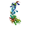
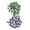
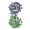
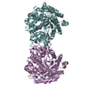
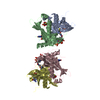
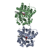
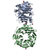
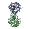
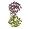
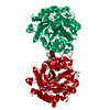
 STREPTOCOCCUS PNEUMONIAE (bacteria)
 X-RAY DIFFRACTION /
  Authors
 Citation
 Structure visualization
 Downloads & links
 Other downloads
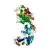

 PDBj
PDBj

 Assembly

 Mass: 65.409 Da / Num. of mol.: 9 / Source method: obtained synthetically / Formula: Zn
 Mass: 18.015 Da / Num. of mol.: 570 / Source method: isolated from a natural source / Formula: H2O
 Sample preparation
 Processing