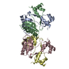
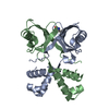
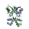
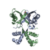
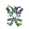
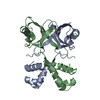
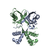
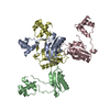
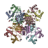
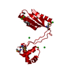
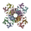
Entry Database : PDB / ID : 2w25Title Crystal structure of Glu104Ala mutant PROBABLE TRANSCRIPTIONAL REGULATORY PROTEIN Keywords / / / / / / Function / homology Function Domain/homology Component
/ / / / / / / / / / / / / / / / / / / / / / / / / / / / / / / Biological species MYCOBACTERIUM TUBERCULOSIS (bacteria)Method / / Resolution : 2.15 Å Authors Shrivastava, T. / RAmachandran, R. Journal : J.Mol.Biol. / Year : 2009Title : Ligand-Induced Structural Transitions, Mutational Analysis, and 'Open' Quaternary Structure of the M. Tuberculosis Feast/Famine Regulatory Protein (Rv3291C).Authors : Shrivastava, T. / Dey, A. / Ramachandran, R. History Deposition Oct 24, 2008 Deposition site / Processing site Revision 1.0 Nov 17, 2009 Provider / Type Revision 1.1 May 8, 2011 Group Revision 1.2 Jul 13, 2011 Group Revision 1.3 Oct 16, 2019 Group / Experimental preparation / OtherCategory / pdbx_database_status / reflns_shellItem _exptl_crystal_grow.method / _exptl_crystal_grow.pdbx_details ... _exptl_crystal_grow.method / _exptl_crystal_grow.pdbx_details / _exptl_crystal_grow.temp / _pdbx_database_status.status_code_sf / _reflns_shell.Rmerge_I_obs Revision 1.4 Dec 13, 2023 Group / Database references / Refinement descriptionCategory chem_comp_atom / chem_comp_bond ... chem_comp_atom / chem_comp_bond / database_2 / pdbx_initial_refinement_model Item / _database_2.pdbx_database_accession
Show all Show less Remark 700 SHEET DETERMINATION METHOD: DSSP THE SHEETS PRESENTED AS "AB" IN EACH CHAIN ON SHEET RECORDS BELOW ... SHEET DETERMINATION METHOD: DSSP THE SHEETS PRESENTED AS "AB" IN EACH CHAIN ON SHEET RECORDS BELOW IS ACTUALLY AN 8-STRANDED BARREL THIS IS REPRESENTED BY A 9-STRANDED SHEET IN WHICH THE FIRST AND LAST STRANDS ARE IDENTICAL.
 Movie
Movie Controller


 Open data
 Basic information
 Components
 Keywords
 Function and homology information
 MYCOBACTERIUM TUBERCULOSIS (bacteria)
 X-RAY DIFFRACTION /
  Authors
 Citation
 Structure visualization
 Downloads & links
 Other downloads
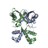


 PDBj
PDBj Assembly

 Mass: 18.015 Da / Num. of mol.: 72 / Source method: isolated from a natural source / Formula: H2O
 Sample preparation
 Processing