- PDB-2ivm: Crystal structure of a transcriptional regulator -
+
Open data
ID or keywords:
Loading...
-
Basic information
Entry
Database: PDB / ID: 2ivm
Title
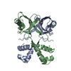
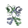
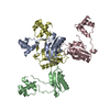
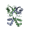
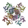
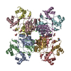
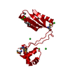
Crystal structure of a transcriptional regulator
Components
TRANSCRIPTIONAL REGULATORY PROTEIN
Keywords
TRANSCRIPTION / TRANSCRIPTIONAL REGULATOR
Function / homology
Function and homology information
amino acid binding / response to amino acid / protein homooligomerization / sequence-specific DNA binding / DNA binding / cytosol Similarity search - Function
SHEET DETERMINATION METHOD: DSSP THE SHEETS PRESENTED AS "AB" IN EACH CHAIN ON SHEET RECORDS BELOW ... SHEET DETERMINATION METHOD: DSSP THE SHEETS PRESENTED AS "AB" IN EACH CHAIN ON SHEET RECORDS BELOW IS ACTUALLY AN 8-STRANDED BARREL THIS IS REPRESENTED BY A 9-STRANDED SHEET IN WHICH THE FIRST AND LAST STRANDS ARE IDENTICAL.
 Movie
Movie Controller


 Open data
 Basic information
 Components
 Keywords
 Function and homology information
 MYCOBACTERIUM TUBERCULOSIS (bacteria)
 X-RAY DIFFRACTION /
  Authors
 Citation
 Structure visualization
 Downloads & links
 Other downloads
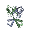


 PDBj
PDBj Assembly
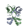

 Mass: 18.015 Da / Num. of mol.: 40 / Source method: isolated from a natural source / Formula: H2O
 Sample preparation
 Processing