 Movie
Movie Controller

[English] 日本語
 Yorodumi
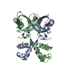
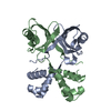
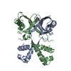
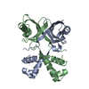
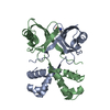
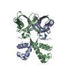
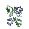
- PDB-2vbw: Feast or famine regulatory protein (Rv3291c)from M. tuberculosis ... -
+ Open data

- Basic information
| Entry | Database: PDB / ID: 2vbw | ||||||
|---|---|---|---|---|---|---|---|
| Title | Feast or famine regulatory protein (Rv3291c)from M. tuberculosis complexed with L-Phenylalanine | ||||||
 Components Components | TRANSCRIPTIONAL REGULATORY PROTEIN | ||||||
 Keywords Keywords | DNA BINDING PROTEIN / M. TUBERCULOSIS / PHENYLALANINE COMPLEX / FEAST/FAMINE REGULATORY PROTEIN / DNA-BINDING PROTEIN / TRANSCRIPTION REGULATOR / TRANSCRIPTION REGULATION / LRP / RV3291C / DNA-BINDING / TRANSCRIPTION | ||||||
| Function / homology |  Function and homology information Function and homology informationamino acid binding / response to amino acid / protein homooligomerization / sequence-specific DNA binding / DNA binding / cytosol Similarity search - Function | ||||||
| Biological species |   MYCOBACTERIUM TUBERCULOSIS (bacteria) MYCOBACTERIUM TUBERCULOSIS (bacteria) | ||||||
| Method |  X-RAY DIFFRACTION / MIRAS / Resolution: 2.2 Å X-RAY DIFFRACTION / MIRAS / Resolution: 2.2 Å | ||||||
 Authors Authors | Shrivastava, T. / Ramachandran, R. | ||||||
 Citation Citation | Journal: Nucleic Acids Res. / Year: 2007 Title: Mechanistic Insights from the Crystal Structures of a Feast/Famine Regulatory Protein from Mycobacterium Tuberculosis H37Rv. Authors: Shrivastava, T. / Ramachandran, R. | ||||||
| History |
| ||||||
| Remark 700 | SHEET DETERMINATION METHOD: DSSP THE SHEETS PRESENTED AS "AB" IN EACH CHAIN ON SHEET RECORDS BELOW ... SHEET DETERMINATION METHOD: DSSP THE SHEETS PRESENTED AS "AB" IN EACH CHAIN ON SHEET RECORDS BELOW IS ACTUALLY AN 8-STRANDED BARREL THIS IS REPRESENTED BY A 9-STRANDED SHEET IN WHICH THE FIRST AND LAST STRANDS ARE IDENTICAL. |
- Structure visualization
| Structure viewer | Molecule: MolmilJmol/JSmol |
|---|
- Downloads & links
-Download
| PDBx/mmCIF format | 2vbw.cif.gz | 68.1 KB | Display | PDBx/mmCIF format |
|---|---|---|---|---|
| PDB format | pdb2vbw.ent.gz | 52.4 KB | Display | PDB format |
| PDBx/mmJSON format | 2vbw.json.gz | Tree view | PDBx/mmJSON format | |
| Others |  Other downloads Other downloads |
-Validation report
| Arichive directory | https://data.pdbj.org/pub/pdb/validation_reports/vb/2vbwftp://data.pdbj.org/pub/pdb/validation_reports/vb/2vbw | HTTPS FTP |
|---|
-Related structure data
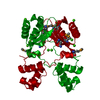
| Related structure data |  2ivmC  2vbxC 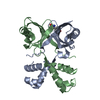 2vbyC 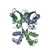 2vbzC 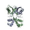 2vc0C  2vc1C C: citing same article ( |
|---|---|
| Similar structure data |

-Links
 PDBj
PDBj


- Assembly
| Deposited unit | 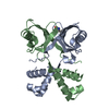
| ||||||||
|---|---|---|---|---|---|---|---|---|---|
| 1 |
| ||||||||
| Unit cell |
| ||||||||
| Components on special symmetry positions |
|
-Components
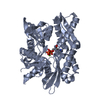
| #1: Protein | Mass: 16530.596 Da / Num. of mol.: 2 Source method: isolated from a genetically manipulated source Details: RV3291C COMPLEXED TO L-PHENYLALANINE / Source: (gene. exp.) MYCOBACTERIUM TUBERCULOSIS (bacteria) / Strain: H37RV / Production host: #2: Chemical | ChemComp-PHE / |   Type: L-peptide linking / Mass: 165.189 Da / Num. of mol.: 1 / Source method: obtained synthetically / Formula: C9H11NO2 Type: L-peptide linking / Mass: 165.189 Da / Num. of mol.: 1 / Source method: obtained synthetically / Formula: C9H11NO2#3: Water | ChemComp-HOH / |  Mass: 18.015 Da / Num. of mol.: 105 / Source method: isolated from a natural source / Formula: H2O Mass: 18.015 Da / Num. of mol.: 105 / Source method: isolated from a natural source / Formula: H2O |
|---|
-Experimental details
-Experiment
| Experiment | Method: X-RAY DIFFRACTION / Number of used crystals: 1 |
|---|
- Sample preparation
| Crystal | Density Matthews: 3.84 Å3/Da / Density % sol: 67.68 % / Description: NONE |
|---|
-Data collection
| Diffraction | Mean temperature: 298 K |
|---|---|
| Diffraction source | Source: ROTATING ANODE / Type: RIGAKU RU300 / Wavelength: 1.5418 |
| Detector | Type: MARRESEARCH / Detector: AREA DETECTOR / Details: MIRRORS |
| Radiation | Protocol: SINGLE WAVELENGTH / Monochromatic (M) / Laue (L): M / Scattering type: x-ray |
| Radiation wavelength | Wavelength: 1.5418 Å / Relative weight: 1 |
| Reflection | Resolution: 2.2→20 Å / Num. obs: 26683 / % possible obs: 99.9 % / Observed criterion σ(I): 2 / Redundancy: 5.1 % / Rmerge(I) obs: 0.1 / Net I/σ(I): 11.9 |
| Reflection shell | Resolution: 2.2→2.32 Å / Redundancy: 5.1 % / Rmerge(I) obs: 0.76 / Mean I/σ(I) obs: 2.2 / % possible all: 100 |
- Processing
| Software |
| ||||||||||||||||||||
|---|---|---|---|---|---|---|---|---|---|---|---|---|---|---|---|---|---|---|---|---|---|
| Refinement | Method to determine structure: MIRAS Starting model: NONE Resolution: 2.2→71.43 Å / Cor.coef. Fo:Fc: 0.939 / Cor.coef. Fo:Fc free: 0.918 / Cross valid method: THROUGHOUT / ESU R: 0.144 / ESU R Free: 0.173 / Stereochemistry target values: MAXIMUM LIKELIHOOD Details: HYDROGENS HAVE BEEN ADDED IN THE RIDING POSITIONS. RESIDUES 1-3 ARE DISORDERED IN THE STRUCTURE
| ||||||||||||||||||||
| Solvent computation | Ion probe radii: 0.8 Å / Shrinkage radii: 0.8 Å / VDW probe radii: 1.2 Å / Solvent model: MASK | ||||||||||||||||||||
| Displacement parameters | Biso mean: 40.77 Å2
| ||||||||||||||||||||
| Refinement step | Cycle: LAST / Resolution: 2.2→71.43 Å
| ||||||||||||||||||||
| LS refinement shell | Resolution: 2.2→2.26 Å / Total num. of bins used: 20 /
|
