 Movie
Movie Controller

[English] 日本語
 Yorodumi
- PDB-2pn6: Crystal Structure of S32A of ST1022-Gln complex from Sulfolobus t... -
+ Open data

- Basic information
| Entry | Database: PDB / ID: 2pn6 | ||||||
|---|---|---|---|---|---|---|---|
| Title | Crystal Structure of S32A of ST1022-Gln complex from Sulfolobus tokodaii | ||||||
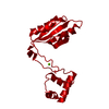
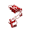
 Components Components | 150aa long hypothetical transcriptional regulator | ||||||
 Keywords Keywords | TRANSCRIPTION / Transcriptional regulator / Lrp/AsnC family Gln binding / ST1022 / Structural Genomics / NPPSFA / National Project on Protein Structural and Functional Analyses / RIKEN Structural Genomics/Proteomics Initiative / RSGI | ||||||
| Function / homology |  Function and homology information Function and homology informationresponse to amino acid / sequence-specific DNA binding / DNA-templated transcription / cytosol Similarity search - Function | ||||||
| Biological species |   Sulfolobus tokodaii (archaea) Sulfolobus tokodaii (archaea) | ||||||
| Method |  X-RAY DIFFRACTION / SYNCHROTRON / MOLECULAR REPLACEMENT / Resolution: 1.44 Å X-RAY DIFFRACTION / SYNCHROTRON / MOLECULAR REPLACEMENT / Resolution: 1.44 Å | ||||||
 Authors Authors | Kumarevel, T.S. / Karthe, P. / Nakano, N. / Shinkai, A. / Yokoyama, S. / RIKEN Structural Genomics/Proteomics Initiative (RSGI) | ||||||
 Citation Citation | Journal: Nucleic Acids Res. / Year: 2008 Title: Crystal structure of glutamine receptor protein from Sulfolobus tokodaii strain 7 in complex with its effector L-glutamine: implications of effector binding in molecular association and DNA binding Authors: Kumarevel, T.S. / Nakano, N. / Ponnuraj, K. / Gopinath, S.C. / Sakamoto, K. / Shinkai, A. / Kumar, P.K. / Yokoyama, S. | ||||||
| History |
|
- Structure visualization
| Structure viewer | Molecule: MolmilJmol/JSmol |
|---|
- Downloads & links
-Download
| PDBx/mmCIF format | 2pn6.cif.gz | 50 KB | Display | PDBx/mmCIF format |
|---|---|---|---|---|
| PDB format | pdb2pn6.ent.gz | 35 KB | Display | PDB format |
| PDBx/mmJSON format | 2pn6.json.gz | Tree view | PDBx/mmJSON format | |
| Others |  Other downloads Other downloads |
-Validation report
| Arichive directory | https://data.pdbj.org/pub/pdb/validation_reports/pn/2pn6ftp://data.pdbj.org/pub/pdb/validation_reports/pn/2pn6 | HTTPS FTP |
|---|
-Related structure data
| Related structure data |  2e7wSC  2e7xC  2efnC  2efoC  2efpC  2efqC  2pmhC  2yx4C  2yx7C S: Starting model for refinement C: citing same article ( |
|---|---|
| Similar structure data | |
| Other databases |


-Links
 PDBj
PDBj- Assembly
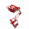
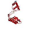
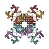
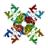
| Deposited unit | 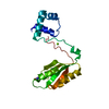
| ||||||||
|---|---|---|---|---|---|---|---|---|---|
| 1 | x 8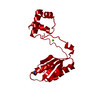
| ||||||||
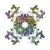
| Unit cell |
|
-Components
| #1: Protein | Mass: 17482.473 Da / Num. of mol.: 1 / Mutation: S32A Source method: isolated from a genetically manipulated source Source: (gene. exp.) Sulfolobus tokodaii (archaea) / Gene: ST1022 / Plasmid: BL21(DE3) / Production host:  |
|---|---|
| #2: Chemical | ChemComp-MG /   Mass: 24.305 Da / Num. of mol.: 1 / Source method: obtained synthetically / Formula: Mg Mass: 24.305 Da / Num. of mol.: 1 / Source method: obtained synthetically / Formula: Mg |
| #3: Chemical | ChemComp-GLN /   Type: L-peptide linking / Mass: 146.144 Da / Num. of mol.: 1 / Source method: obtained synthetically / Formula: C5H10N2O3 Type: L-peptide linking / Mass: 146.144 Da / Num. of mol.: 1 / Source method: obtained synthetically / Formula: C5H10N2O3 |
| #4: Water | ChemComp-HOH /  Mass: 18.015 Da / Num. of mol.: 195 / Source method: isolated from a natural source / Formula: H2O Mass: 18.015 Da / Num. of mol.: 195 / Source method: isolated from a natural source / Formula: H2O |
-Experimental details
-Experiment
| Experiment | Method: X-RAY DIFFRACTION / Number of used crystals: 1 |
|---|
- Sample preparation
| Crystal | Density Matthews: 2.82 Å3/Da / Density % sol: 56.45 % |
|---|---|
| Crystal grow | Temperature: 293 K / Method: vapor diffusion, sitting drop / pH: 6.2 Details: 20% PPG P400, 30% i-PrOH, 70mM Na3 Citrate, Cymal-7, pH 6.2, VAPOR DIFFUSION, SITTING DROP, temperature 293K |
-Data collection
| Diffraction | Mean temperature: 180 K |
|---|---|
| Diffraction source | Source: SYNCHROTRON / Site: SPring-8  / Beamline: BL26B2 / Wavelength: 1 Å / Beamline: BL26B2 / Wavelength: 1 Å |
| Detector | Type: RIGAKU JUPITER 210 / Detector: CCD / Date: Mar 7, 2007 |
| Radiation | Monochromator: Si / Protocol: SINGLE WAVELENGTH / Monochromatic (M) / Laue (L): M / Scattering type: x-ray |
| Radiation wavelength | Wavelength: 1 Å / Relative weight: 1 |
| Reflection | Resolution: 1.44→50 Å / Num. all: 35897 / Num. obs: 35897 / % possible obs: 98.3 % / Observed criterion σ(F): 2 / Observed criterion σ(I): 2 / Redundancy: 11.5 % / Biso Wilson estimate: 17.8 Å2 / Rmerge(I) obs: 0.059 |
| Reflection shell | Resolution: 1.44→1.49 Å / Redundancy: 3.5 % / Rmerge(I) obs: 0.49 / Num. unique all: 3013 / % possible all: 84.5 |
- Processing
| Software |
| ||||||||||||||||||||||||||||||||||||
|---|---|---|---|---|---|---|---|---|---|---|---|---|---|---|---|---|---|---|---|---|---|---|---|---|---|---|---|---|---|---|---|---|---|---|---|---|---|
| Refinement | Method to determine structure: MOLECULAR REPLACEMENT Starting model: 2E7W Resolution: 1.44→19.99 Å / Rfactor Rfree error: 0.005 / Data cutoff high absF: 1949192.02 / Data cutoff low absF: 0 / Isotropic thermal model: RESTRAINED / Cross valid method: THROUGHOUT / σ(F): 0 / Stereochemistry target values: Engh & Huber
| ||||||||||||||||||||||||||||||||||||
| Solvent computation | Solvent model: FLAT MODEL / Bsol: 49.1207 Å2 / ksol: 0.368202 e/Å3 | ||||||||||||||||||||||||||||||||||||
| Displacement parameters | Biso mean: 21.7 Å2
| ||||||||||||||||||||||||||||||||||||
| Refine analyze |
| ||||||||||||||||||||||||||||||||||||
| Refinement step | Cycle: LAST / Resolution: 1.44→19.99 Å
| ||||||||||||||||||||||||||||||||||||
| Refine LS restraints |
| ||||||||||||||||||||||||||||||||||||
| LS refinement shell | Resolution: 1.44→1.53 Å / Rfactor Rfree error: 0.019 / Total num. of bins used: 6
| ||||||||||||||||||||||||||||||||||||
| Xplor file |
|
