 Movie
Movie Controller

[English] 日本語
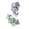
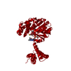
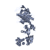
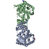
 Yorodumi
- PDB-2vsg: A Structural Motif in the Variant Surface Glycoproteins of Trypan... -
+ Open data

- Basic information
| Entry | Database: PDB / ID: 2vsg | ||||||
|---|---|---|---|---|---|---|---|
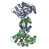
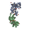
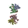
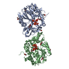
| Title | A Structural Motif in the Variant Surface Glycoproteins of Trypanosoma Brucei | ||||||
 Components Components | VARIANT SURFACE GLYCOPROTEIN ILTAT 1.24 | ||||||
 Keywords Keywords | MEMBRANE PROTEIN / VSG / TRYPANOSOME / ANTIGENIC VARIATION | ||||||
| Function / homology |  Function and homology information Function and homology informationsymbiont-mediated evasion of host immune response / side of membrane / plasma membrane Similarity search - Function | ||||||
| Biological species |  | ||||||
| Method |  X-RAY DIFFRACTION / SIRAS / Resolution: 2.7 Å X-RAY DIFFRACTION / SIRAS / Resolution: 2.7 Å | ||||||
 Authors Authors | Blum, M.L. / Down, J.A. / Metcalf, P. / Freymann, D.M. / Wiley, D.C. | ||||||
 Citation Citation | Journal: Nature / Year: 1993 Title: A structural motif in the variant surface glycoproteins of Trypanosoma brucei. Authors: Blum, M.L. / Down, J.A. / Gurnett, A.M. / Carrington, M. / Turner, M.J. / Wiley, D.C. #1: Journal: Thesis / Year: 1990Title: The Structure of a Vsg Variable Domain from Trypanosoma Brucei at 2.7 A Resolution Authors: Blum, M.L. #2: Journal: J.Biol.Chem. / Year: 1988Title: Crystallization of Amino-Terminal Domains and Domain Fragments of Variant Surface Glycoproteins from Trypanosoma Brucei Brucei Authors: Metcalf, P. / Down, J.A. / Turner, M. / Wiley, D.C. #3: Journal: Nature / Year: 1987Title: Two Variant Surface Glycoproteins of Trypanosoma Brucei of Diffrent Sequence Classes Have Similar 6 Angstrom Resolution X-Ray Structures Authors: Metcalf, P. / Blum, M. / Freymann, D. / Turner, M. / Wiley, D.C. | ||||||
| History |
|
- Structure visualization
| Structure viewer | Molecule: MolmilJmol/JSmol |
|---|
- Downloads & links
-Download
| PDBx/mmCIF format | 2vsg.cif.gz | 142.5 KB | Display | PDBx/mmCIF format |
|---|---|---|---|---|
| PDB format | pdb2vsg.ent.gz | 113.1 KB | Display | PDB format |
| PDBx/mmJSON format | 2vsg.json.gz | Tree view | PDBx/mmJSON format | |
| Others |  Other downloads Other downloads |
-Validation report
| Arichive directory | https://data.pdbj.org/pub/pdb/validation_reports/vs/2vsgftp://data.pdbj.org/pub/pdb/validation_reports/vs/2vsg | HTTPS FTP |
|---|
-Related structure data
| Related structure data | |
|---|---|
| Similar structure data |


-Links
 PDBj
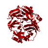
PDBj- Assembly
| Deposited unit | 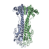
| ||||||||||
|---|---|---|---|---|---|---|---|---|---|---|---|
| 1 |
| ||||||||||
| Unit cell |
| ||||||||||
| Noncrystallographic symmetry (NCS) | NCS oper: (Code: given Matrix: (-0.991703, 0.074356, 0.104862), Vector: |
-Components
| #1: Protein | Mass: 38286.742 Da / Num. of mol.: 2 / Fragment: N-TERMINAL VARIABLE DOMAIN / Source method: isolated from a natural source / Source: (natural) #2: Water | ChemComp-HOH / |  Mass: 18.015 Da / Num. of mol.: 83 / Source method: isolated from a natural source / Formula: H2O Mass: 18.015 Da / Num. of mol.: 83 / Source method: isolated from a natural source / Formula: H2OHas protein modification | Y | |
|---|
-Experimental details
-Experiment
| Experiment | Method: X-RAY DIFFRACTION / Number of used crystals: 7 |
|---|
- Sample preparation
| Crystal | Density Matthews: 3.07 Å3/Da / Density % sol: 59.96 % Description: PHASES TO 4.2 A WERE OBTAINED BY SIRAS, IMPROVED BY NCS AVERAGING, AND EXTENDED TO 3.8 A. A PARTIAL MODEL WAS BUILT, AND RESOLUTION EXTENDED TO 2.7 A BY NCS AVERAGING AND PHASE COMBINATION. |
|---|---|
| Crystal grow | pH: 6.5 / Details: pH 6.5 |
| Crystal grow | *PLUS Method: unknown |
-Data collection
| Diffraction | Mean temperature: 277 K |
|---|---|
| Diffraction source | Wavelength: 1.5418 |
| Detector | Type: XENTRONICS / Detector: AREA DETECTOR / Date: Aug 5, 1986 / Details: MIRRORS |
| Radiation | Protocol: SINGLE WAVELENGTH / Monochromatic (M) / Laue (L): M / Scattering type: x-ray |
| Radiation wavelength | Wavelength: 1.5418 Å / Relative weight: 1 |
| Reflection | Resolution: 2.7→10 Å / Num. all: 24682 / Num. obs: 24682 / % possible obs: 93 % / Observed criterion σ(I): 0 / Redundancy: 4.4 % / Rsym value: 0.102 |
| Reflection shell | Resolution: 2.7→2.83 Å / Redundancy: 3.64 % / Rsym value: 0.39 |
| Reflection | *PLUS Num. measured all: 107903 / Rmerge(I) obs: 0.102 |
| Reflection shell | *PLUS Rmerge(I) obs: 0.39 |
- Processing
| Software |
| ||||||||||||||||||||||||||||||||||||||||||||||||||||||||||||
|---|---|---|---|---|---|---|---|---|---|---|---|---|---|---|---|---|---|---|---|---|---|---|---|---|---|---|---|---|---|---|---|---|---|---|---|---|---|---|---|---|---|---|---|---|---|---|---|---|---|---|---|---|---|---|---|---|---|---|---|---|---|
| Refinement | Method to determine structure: SIRAS / Resolution: 2.7→6 Å / Isotropic thermal model: RESTRAINED / σ(F): 2 Details: SEE REMARK 6 THIS STRUCTURE WAS REFINED BEFORE THE USE OF THE FREE R WAS INTRODUCED. IT IS MISSING SOME OF THE QUALITY INDICATORS NOW WIDELY USED. RELATIVELY LARGE DEVIATIONS FROM STANDARD ...Details: SEE REMARK 6 THIS STRUCTURE WAS REFINED BEFORE THE USE OF THE FREE R WAS INTRODUCED. IT IS MISSING SOME OF THE QUALITY INDICATORS NOW WIDELY USED. RELATIVELY LARGE DEVIATIONS FROM STANDARD GEOMETRY CAN BE ASCRIBED TO BOTH THE LIMITED RESOLUTION OF THE DATA AND THE STATE OF THE ART WHEN THE STRUCTURE WAS REFINED, AND MAY BE INDICATIVE OF SOME OVERFITTING. NEVERTHELESS THE ELECTRON DENSITY MAP WAS WELL DEFINED AND ALLOWED, FOR EXAMPLE, IDENTIFICATION OF THREE SEQUENCING ERRORS WHICH WERE SUBSEQUENTLY VERIFIED.
| ||||||||||||||||||||||||||||||||||||||||||||||||||||||||||||
| Displacement parameters | Biso mean: 34.2 Å2 | ||||||||||||||||||||||||||||||||||||||||||||||||||||||||||||
| Refinement step | Cycle: LAST / Resolution: 2.7→6 Å
| ||||||||||||||||||||||||||||||||||||||||||||||||||||||||||||
| Refine LS restraints |
| ||||||||||||||||||||||||||||||||||||||||||||||||||||||||||||
| Refine LS restraints NCS | NCS model details: RESTRAINTS |
