 Movie
Movie Controller

[English] 日本語
 Yorodumi
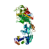
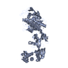
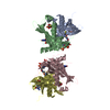
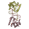
- PDB-1vsg: 2.9 ANGSTROMS RESOLUTION STRUCTURE OF THE N-TERMINAL DOMAIN OF A ... -
+ Open data

- Basic information
| Entry | Database: PDB / ID: 1vsg | |||||||||
|---|---|---|---|---|---|---|---|---|---|---|
| Title | 2.9 ANGSTROMS RESOLUTION STRUCTURE OF THE N-TERMINAL DOMAIN OF A VARIANT SURFACE GLYCOPROTEIN FROM TRYPANOSOMA BRUCEI | |||||||||
 Components Components | VARIANT SURFACE GLYCOPROTEIN MITAT 1.2 | |||||||||
 Keywords Keywords | GLYCOPROTEIN | |||||||||
| Function / homology |  Function and homology information Function and homology informationsymbiont-mediated evasion of host immune response / side of membrane / plasma membrane Similarity search - Function | |||||||||
| Biological species |  | |||||||||
| Method |  X-RAY DIFFRACTION / Resolution: 2.9 Å X-RAY DIFFRACTION / Resolution: 2.9 Å | |||||||||
 Authors Authors | Freymann, D. / Down, J. / Wiley, D.C. | |||||||||
 Citation Citation | Journal: J.Mol.Biol. / Year: 1990 Title: 2.9 A resolution structure of the N-terminal domain of a variant surface glycoprotein from Trypanosoma brucei. Authors: Freymann, D. / Down, J. / Carrington, M. / Roditi, I. / Turner, M. / Wiley, D. #1: Journal: Nature / Year: 1987Title: Two Variant Surface Gylcoproteins of Trypanosoma Brucei of Different Sequence Classes Have Similar 6 Angstroms Resolution X-Ray Structures Authors: Metcalf, P. / Blum, M. / Freymann, D. / Turner, M. / Wiley, D.C. #2: Journal: Nature / Year: 1984Title: 6 Angstroms-Resolution X-Ray Structure of a Variable Surface Glycoprotein from Trypanosoma Brucei Authors: Freymann, D.M. / Metcalf, P. / Turner, M. / Wiley, D.C. | |||||||||
| History |
|
- Structure visualization
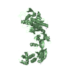
| Structure viewer | Molecule: MolmilJmol/JSmol |
|---|
- Downloads & links
-Download
| PDBx/mmCIF format | 1vsg.cif.gz | 145.4 KB | Display | PDBx/mmCIF format |
|---|---|---|---|---|
| PDB format | pdb1vsg.ent.gz | 114.6 KB | Display | PDB format |
| PDBx/mmJSON format | 1vsg.json.gz | Tree view | PDBx/mmJSON format | |
| Others |  Other downloads Other downloads |
-Validation report
| Arichive directory | https://data.pdbj.org/pub/pdb/validation_reports/vs/1vsgftp://data.pdbj.org/pub/pdb/validation_reports/vs/1vsg | HTTPS FTP |
|---|
-Related structure data
| Similar structure data |
|---|

-Links
 PDBj
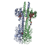
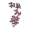
PDBj- Assembly
| Deposited unit | 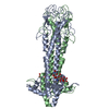
| ||||||||
|---|---|---|---|---|---|---|---|---|---|
| 1 |
| ||||||||
| Unit cell |
| ||||||||
| Atom site foot note | 1: RESIDUES ASN 183 - ASP 184, THR 265 - ALA 266, LYS 285 -GLU 289, AND ASP 328 - ASN 329 IN BOTH THE A CHAIN AND B CHAIN ARE POORLY DEFINED IN THE ELECTRON DENSITY MAP. | ||||||||
| Noncrystallographic symmetry (NCS) | NCS oper: (Code: given Matrix: (-0.06906, 0.19176, 0.97901), Vector: Details | THE TRANSFORMATION PRESENTED ON THE *MTRIX* RECORDS BELOW WILL YIELD APPROXIMATE COORDINATES FOR CHAIN *B* WHEN APPLIED TO CHAIN *A*. | |
-Components
| #1: Protein | Mass: 38844.660 Da / Num. of mol.: 2 Source method: isolated from a genetically manipulated source Source: (gene. exp.) #2: Polysaccharide | beta-D-mannopyranose-(1-4)-2-acetamido-2-deoxy-beta-D-glucopyranose-(1-4)-2-acetamido-2-deoxy-beta- ...beta-D-mannopyranose-(1-4)-2-acetamido-2-deoxy-beta-D-glucopyranose-(1-4)-2-acetamido-2-deoxy-beta-D-glucopyranose | Source method: isolated from a genetically manipulated source #3: Polysaccharide | alpha-D-mannopyranose-(1-4)-2-acetamido-2-deoxy-beta-D-glucopyranose-(1-4)-2-acetamido-2-deoxy-beta- ...alpha-D-mannopyranose-(1-4)-2-acetamido-2-deoxy-beta-D-glucopyranose-(1-4)-2-acetamido-2-deoxy-beta-D-glucopyranose | Source method: isolated from a genetically manipulated source #4: Water | ChemComp-HOH / |  Mass: 18.015 Da / Num. of mol.: 18 / Source method: isolated from a natural source / Formula: H2O Mass: 18.015 Da / Num. of mol.: 18 / Source method: isolated from a natural source / Formula: H2OHas protein modification | Y | |
|---|
-Experimental details
-Experiment
| Experiment | Method: X-RAY DIFFRACTION |
|---|
- Sample preparation
| Crystal | Density Matthews: 3.71 Å3/Da / Density % sol: 66.81 % | ||||||||||||||||||||||||||||||||||||||||||
|---|---|---|---|---|---|---|---|---|---|---|---|---|---|---|---|---|---|---|---|---|---|---|---|---|---|---|---|---|---|---|---|---|---|---|---|---|---|---|---|---|---|---|---|
| Crystal grow | *PLUS pH: 7 / Method: vapor diffusion, hanging drop | ||||||||||||||||||||||||||||||||||||||||||
| Components of the solutions | *PLUS
|
-Data collection
| Reflection | *PLUS Highest resolution: 2.9 Å / Num. obs: 24767 / Num. measured all: 56659 / Rmerge(I) obs: 0.087 |
|---|
- Processing
| Software | Name: X-PLOR / Classification: refinement | ||||||||||||||||||||||||||||||||||||||||||||||||||||||||||||
|---|---|---|---|---|---|---|---|---|---|---|---|---|---|---|---|---|---|---|---|---|---|---|---|---|---|---|---|---|---|---|---|---|---|---|---|---|---|---|---|---|---|---|---|---|---|---|---|---|---|---|---|---|---|---|---|---|---|---|---|---|---|
| Refinement | Rfactor Rwork: 0.22 / Highest resolution: 2.9 Å Details: RESIDUES ASN 183 - ASP 184, THR 265 - ALA 266, LYS 285 -GLU 289, AND ASP 328 - ASN 329 IN BOTH THE A CHAIN AND B CHAIN ARE POORLY DEFINED IN THE ELECTRON DENSITY MAP | ||||||||||||||||||||||||||||||||||||||||||||||||||||||||||||
| Refinement step | Cycle: LAST / Highest resolution: 2.9 Å
| ||||||||||||||||||||||||||||||||||||||||||||||||||||||||||||
| Refine LS restraints |
| ||||||||||||||||||||||||||||||||||||||||||||||||||||||||||||
| Refinement | *PLUS Highest resolution: 2.9 Å / Rfactor obs: 0.22 / Lowest resolution: 7 Å / Num. reflection obs: 5510 | ||||||||||||||||||||||||||||||||||||||||||||||||||||||||||||
| Solvent computation | *PLUS | ||||||||||||||||||||||||||||||||||||||||||||||||||||||||||||
| Displacement parameters | *PLUS | ||||||||||||||||||||||||||||||||||||||||||||||||||||||||||||
| Refine LS restraints | *PLUS
|
