 Movie
Movie Controller


+ Open data

- Basic information
| Entry | Database: PDB / ID: 2vp8 | ||||||
|---|---|---|---|---|---|---|---|
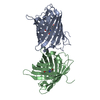
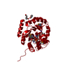
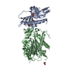
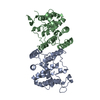
| Title | Structure of Mycobacterium tuberculosis Rv1207 | ||||||
 Components Components | DIHYDROPTEROATE SYNTHASE 2 | ||||||
 Keywords Keywords | TRANSFERASE / DIHYDROPTEROATE SYNTHASE / MYCOBACTERIUM TUBERCULOSIS / RV1207 / FOLATE BIOSYNTHESIS / ANTIBIOTIC RESISTANCE | ||||||
| Function / homology |  Function and homology information Function and homology informationfolic acid-containing compound biosynthetic process / dihydropteroate synthase activity / cytosol Similarity search - Function | ||||||
| Biological species |   MYCOBACTERIUM TUBERCULOSIS (bacteria) MYCOBACTERIUM TUBERCULOSIS (bacteria) | ||||||
| Method |  X-RAY DIFFRACTION / SYNCHROTRON / MOLECULAR REPLACEMENT / Resolution: 2.64 Å X-RAY DIFFRACTION / SYNCHROTRON / MOLECULAR REPLACEMENT / Resolution: 2.64 Å | ||||||
 Authors Authors | Gengenbacher, M. / Xu, T. / Niyomwattanakit, P. / Spraggon, G. / Dick, T. | ||||||
 Citation Citation | Journal: Fems Microbiol.Lett. / Year: 2008 Title: Biochemical and Structural Characterization of the Putative Dihydropteroate Synthase Ortholog Rv1207 of Mycobacterium Tuberculosis. Authors: Gengenbacher, M. / Xu, T. / Niyomwattanakit, P. / Spraggon, G. / Dick, T. #1: Journal: J.Mol.Biol. / Year: 2000Title: Crystal Structure of Mycobacterium Tuberculosis 6-Hydroxymethyl-7,8-Dihydropteroate Synthase in Complex with Pterin Monophosphate: New Insight Into the Enzymatic Mechanism and Sulfa-Drug Action Authors: Baca, A.M. / Sirawaraporn, R. / Turley, S. / Sirawaraporn, W. / Hol, W.G.J. | ||||||
| History |
|
- Structure visualization
| Structure viewer | Molecule: MolmilJmol/JSmol |
|---|
- Downloads & links
-Download
| PDBx/mmCIF format | 2vp8.cif.gz | 98.8 KB | Display | PDBx/mmCIF format |
|---|---|---|---|---|
| PDB format | pdb2vp8.ent.gz | 75.4 KB | Display | PDB format |
| PDBx/mmJSON format | 2vp8.json.gz | Tree view | PDBx/mmJSON format | |
| Others |  Other downloads Other downloads |
-Validation report
| Summary document | 2vp8_validation.pdf.gz | 449.3 KB | Display | wwPDB validaton report |
|---|---|---|---|---|
| Full document | 2vp8_full_validation.pdf.gz | 456.9 KB | Display | |
| Data in XML | 2vp8_validation.xml.gz | 18.8 KB | Display | |
| Data in CIF | 2vp8_validation.cif.gz | 25.7 KB | Display | |
| Arichive directory | https://data.pdbj.org/pub/pdb/validation_reports/vp/2vp8ftp://data.pdbj.org/pub/pdb/validation_reports/vp/2vp8 | HTTPS FTP |
-Related structure data
| Related structure data |  1eyeS S: Starting model for refinement |
|---|---|
| Similar structure data |


-Links
 PDBj
PDBj- Assembly
| Deposited unit | 
| ||||||||
|---|---|---|---|---|---|---|---|---|---|
| 1 |
| ||||||||
| Unit cell |
| ||||||||
| Noncrystallographic symmetry (NCS) | NCS oper: (Code: given Matrix: (-0.34353, -0.84941, 0.40061), Vector: |
-Components
| #1: Protein | Mass: 33053.656 Da / Num. of mol.: 2 / Fragment: RESIDUES 2-318 Source method: isolated from a genetically manipulated source Source: (gene. exp.) MYCOBACTERIUM TUBERCULOSIS (bacteria) / Strain: H37RV / Plasmid: PET-21A / Production host: References: UniProt: P64139, UniProt: P9WNC9*PLUS, EC: 2.1.5.15 #2: Chemical | ChemComp-EDO /   Mass: 62.068 Da / Num. of mol.: 4 / Source method: obtained synthetically / Formula: C2H6O2 Mass: 62.068 Da / Num. of mol.: 4 / Source method: obtained synthetically / Formula: C2H6O2#3: Water | ChemComp-HOH / |  Mass: 18.015 Da / Num. of mol.: 55 / Source method: isolated from a natural source / Formula: H2O Mass: 18.015 Da / Num. of mol.: 55 / Source method: isolated from a natural source / Formula: H2O |
|---|
-Experimental details
-Experiment
| Experiment | Method: X-RAY DIFFRACTION |
|---|
- Sample preparation
| Crystal | Density Matthews: 4.13 Å3/Da / Density % sol: 70 % / Description: NONE |
|---|
-Data collection
| Diffraction | Mean temperature: 100 K |
|---|---|
| Diffraction source | Source: SYNCHROTRON / Site: ALS  / Beamline: 5.0.3 / Wavelength: 1 / Beamline: 5.0.3 / Wavelength: 1 |
| Detector | Type: ADSC CCD / Detector: CCD / Date: Feb 27, 2007 |
| Radiation | Protocol: SINGLE WAVELENGTH / Monochromatic (M) / Laue (L): M / Scattering type: x-ray |
| Radiation wavelength | Wavelength: 1 Å / Relative weight: 1 |
| Reflection | Resolution: 2.64→107 Å / Num. obs: 24807 / % possible obs: 99.3 % / Observed criterion σ(I): 0 / Redundancy: 3.2 % / Rmerge(I) obs: 0.07 / Net I/σ(I): 8.5 |
| Reflection shell | Resolution: 2.64→2.8 Å / Redundancy: 3.2 % / Rmerge(I) obs: 0.45 / Mean I/σ(I) obs: 1.6 / % possible all: 99.2 |
- Processing
| Software |
| ||||||||||||||||||||||||||||||||||||||||||||||||||||||||||||||||||||||||||||||||||||||||||||||||||||||||||||||||||||||||||||||||||||||||||||||||||||||||||||||||||||||||||||||||||||||
|---|---|---|---|---|---|---|---|---|---|---|---|---|---|---|---|---|---|---|---|---|---|---|---|---|---|---|---|---|---|---|---|---|---|---|---|---|---|---|---|---|---|---|---|---|---|---|---|---|---|---|---|---|---|---|---|---|---|---|---|---|---|---|---|---|---|---|---|---|---|---|---|---|---|---|---|---|---|---|---|---|---|---|---|---|---|---|---|---|---|---|---|---|---|---|---|---|---|---|---|---|---|---|---|---|---|---|---|---|---|---|---|---|---|---|---|---|---|---|---|---|---|---|---|---|---|---|---|---|---|---|---|---|---|---|---|---|---|---|---|---|---|---|---|---|---|---|---|---|---|---|---|---|---|---|---|---|---|---|---|---|---|---|---|---|---|---|---|---|---|---|---|---|---|---|---|---|---|---|---|---|---|---|---|
| Refinement | Method to determine structure: MOLECULAR REPLACEMENT Starting model: PDB ENTRY 1EYE Resolution: 2.64→19.84 Å / Cor.coef. Fo:Fc: 0.928 / Cor.coef. Fo:Fc free: 0.906 / SU B: 20.695 / SU ML: 0.212 / TLS residual ADP flag: LIKELY RESIDUAL / Cross valid method: THROUGHOUT / ESU R: 0.37 / ESU R Free: 0.279 / Stereochemistry target values: MAXIMUM LIKELIHOOD / Details: HYDROGENS HAVE BEEN ADDED IN THE RIDING POSITIONS.
| ||||||||||||||||||||||||||||||||||||||||||||||||||||||||||||||||||||||||||||||||||||||||||||||||||||||||||||||||||||||||||||||||||||||||||||||||||||||||||||||||||||||||||||||||||||||
| Solvent computation | Ion probe radii: 0.8 Å / Shrinkage radii: 0.8 Å / VDW probe radii: 1.4 Å / Solvent model: MASK | ||||||||||||||||||||||||||||||||||||||||||||||||||||||||||||||||||||||||||||||||||||||||||||||||||||||||||||||||||||||||||||||||||||||||||||||||||||||||||||||||||||||||||||||||||||||
| Displacement parameters | Biso mean: 39.52 Å2
| ||||||||||||||||||||||||||||||||||||||||||||||||||||||||||||||||||||||||||||||||||||||||||||||||||||||||||||||||||||||||||||||||||||||||||||||||||||||||||||||||||||||||||||||||||||||
| Refinement step | Cycle: LAST / Resolution: 2.64→19.84 Å
| ||||||||||||||||||||||||||||||||||||||||||||||||||||||||||||||||||||||||||||||||||||||||||||||||||||||||||||||||||||||||||||||||||||||||||||||||||||||||||||||||||||||||||||||||||||||
| Refine LS restraints |
|