 Movie
Movie Controller


+ Open data

- Basic information
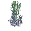
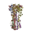
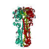
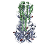
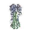
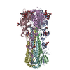
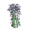
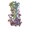
| Entry | Database: PDB / ID: 2viu | |||||||||
|---|---|---|---|---|---|---|---|---|---|---|
| Title | INFLUENZA VIRUS HEMAGGLUTININ | |||||||||
 Components Components | (HEMAGGLUTININ) x 2 | |||||||||
 Keywords Keywords | HEMAGGLUTININ / ENVELOPE PROTEIN / GLYCOPROTEIN | |||||||||
| Function / homology |  Function and homology information Function and homology informationviral budding from plasma membrane / clathrin-dependent endocytosis of virus by host cell / host cell surface receptor binding / fusion of virus membrane with host plasma membrane / fusion of virus membrane with host endosome membrane / viral envelope / virion attachment to host cell / host cell plasma membrane / virion membrane / membrane Similarity search - Function | |||||||||
| Biological species |   Influenza A virus Influenza A virus unidentified influenza virus unidentified influenza virus | |||||||||
| Method |  X-RAY DIFFRACTION / SYNCHROTRON / Resolution: 2.5 Å X-RAY DIFFRACTION / SYNCHROTRON / Resolution: 2.5 Å | |||||||||
 Authors Authors | Bizebard, T. / Fleury, D. / Gigant, B. / Wharton, S.A. / Skehel, J.J. / Knossow, M. | |||||||||
 Citation Citation | Journal: Nat.Struct.Biol. / Year: 1998 Title: Antigen distortion allows influenza virus to escape neutralization. Authors: Fleury, D. / Wharton, S.A. / Skehel, J.J. / Knossow, M. / Bizebard, T. #1: Journal: Nature / Year: 1995Title: Structure of Influenza Virus Haemagglutinin Complexed with a Neutralizing Antibody Authors: Bizebard, T. / Gigant, B. / Rigolet, P. / Rasmussen, B. / Diat, O. / Bosecke, P. / Wharton, S.A. / Skehel, J.J. / Knossow, M. #2: Journal: Acta Crystallogr.,Sect.D / Year: 1994Title: Refined Three-Dimensional Structure of the Fab Fragment of a Murine Iggl, Authors: Bizebard, T. / Daniels, R. / Kahn, R. / Golinelli-Pimpaneau, B. / Skehel, J.J. / Knossow, M. #3: Journal: Nature / Year: 1981Title: Structure of the Haemagglutinin Membrane Glycoprotein of Influenza Virus at 3 A Resolution Authors: Wilson, I.A. / Skehel, J.J. / Wiley, D.C. | |||||||||
| History |
|
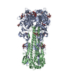
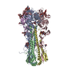
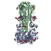
- Structure visualization
| Structure viewer | Molecule: MolmilJmol/JSmol |
|---|
- Downloads & links
-Download
| PDBx/mmCIF format | 2viu.cif.gz | 115.8 KB | Display | PDBx/mmCIF format |
|---|---|---|---|---|
| PDB format | pdb2viu.ent.gz | 88.7 KB | Display | PDB format |
| PDBx/mmJSON format | 2viu.json.gz | Tree view | PDBx/mmJSON format | |
| Others |  Other downloads Other downloads |
-Validation report
| Arichive directory | https://data.pdbj.org/pub/pdb/validation_reports/vi/2viuftp://data.pdbj.org/pub/pdb/validation_reports/vi/2viu | HTTPS FTP |
|---|
-Related structure data


-Links
 PDBj
PDBj


- Assembly
| Deposited unit | 
| ||||||||
|---|---|---|---|---|---|---|---|---|---|
| 1 | 
| ||||||||
| Unit cell |
|
-Components
| #1: Protein | Mass: 36077.512 Da / Num. of mol.: 1 / Mutation: CHAIN A, T131I Source method: isolated from a genetically manipulated source Details: BROMELAIN DIGESTED / Source: (gene. exp.) Influenza A virus (A/X-31(H3N2)) / Genus: Influenzavirus A / Species: Influenza A virus / Strain: X31Description: A REASSORTANT INFLUENZA STRAIN CONTAINING A/AICHI/68 (H3N2) HEMAGGLUTININ References: UniProt: P03437 | ||||
|---|---|---|---|---|---|
| #2: Protein | Mass: 20212.350 Da / Num. of mol.: 1 / Mutation: CHAIN A, T131I Source method: isolated from a genetically manipulated source Details: BROMELAIN DIGESTED / Source: (gene. exp.) unidentified influenza virus / Strain: X31Description: A REASSORTANT INFLUENZA STRAIN CONTAINING A/AICHI/68 (H3N2) HEMAGGLUTININ References: UniProt: P03437 | ||||
| #3: Polysaccharide | beta-D-mannopyranose-(1-4)-2-acetamido-2-deoxy-beta-D-glucopyranose-(1-4)-2-acetamido-2-deoxy-beta- ...beta-D-mannopyranose-(1-4)-2-acetamido-2-deoxy-beta-D-glucopyranose-(1-4)-2-acetamido-2-deoxy-beta-D-glucopyranose Source method: isolated from a genetically manipulated source | ||||
| #4: Sugar | ChemComp-NAG /   Type: D-saccharide, beta linking / Mass: 221.208 Da / Num. of mol.: 5 Type: D-saccharide, beta linking / Mass: 221.208 Da / Num. of mol.: 5Source method: isolated from a genetically manipulated source Formula: C8H15NO6 #5: Water | ChemComp-HOH / |  Mass: 18.015 Da / Num. of mol.: 103 / Source method: isolated from a natural source / Formula: H2O Mass: 18.015 Da / Num. of mol.: 103 / Source method: isolated from a natural source / Formula: H2OHas protein modification | Y | |
-Experimental details
-Experiment
| Experiment | Method: X-RAY DIFFRACTION |
|---|
- Sample preparation
| Crystal | Density Matthews: 4.46 Å3/Da / Density % sol: 66 % | ||||||||||||||||||||||||||||||||||||
|---|---|---|---|---|---|---|---|---|---|---|---|---|---|---|---|---|---|---|---|---|---|---|---|---|---|---|---|---|---|---|---|---|---|---|---|---|---|
| Crystal | *PLUS | ||||||||||||||||||||||||||||||||||||
| Crystal grow | *PLUS Method: vapor diffusion, hanging drop | ||||||||||||||||||||||||||||||||||||
| Components of the solutions | *PLUS
|
-Data collection
| Diffraction source | Source: SYNCHROTRON / Site: LURE  / Beamline: DW32 / Wavelength: 0.97 / Beamline: DW32 / Wavelength: 0.97 |
|---|---|
| Detector | Type: MARRESEARCH / Detector: IMAGE PLATE / Date: Apr 8, 1997 |
| Radiation | Monochromatic (M) / Laue (L): M / Scattering type: x-ray |
| Radiation wavelength | Wavelength: 0.97 Å / Relative weight: 1 |
| Reflection | Resolution: 2.45→20 Å / Num. obs: 36234 / % possible obs: 99.9 % / Redundancy: 4.5 % / Rmerge(I) obs: 0.055 |
| Reflection | *PLUS Num. measured all: 163053 |
- Processing
| Software |
| ||||||||||||||||||||||||||||||||||||||||||||||||||||||||||||
|---|---|---|---|---|---|---|---|---|---|---|---|---|---|---|---|---|---|---|---|---|---|---|---|---|---|---|---|---|---|---|---|---|---|---|---|---|---|---|---|---|---|---|---|---|---|---|---|---|---|---|---|---|---|---|---|---|---|---|---|---|---|
| Refinement | Resolution: 2.5→9 Å / σ(F): 0 Details: THERE IS ONE MONOMER OF THE TRIMERIC HEMAGGLUTININ MOLECULE IN THE ASYMMETRIC UNIT. THE MONOMER OF HEMAGGLUTININ CONSISTS OF TWO CHAINS, IDENTIFIED AS HA1 AND HA2 BY THE THE DEPOSITORS. ...Details: THERE IS ONE MONOMER OF THE TRIMERIC HEMAGGLUTININ MOLECULE IN THE ASYMMETRIC UNIT. THE MONOMER OF HEMAGGLUTININ CONSISTS OF TWO CHAINS, IDENTIFIED AS HA1 AND HA2 BY THE THE DEPOSITORS. CHAINS HA1 AND HA2 HAVE BEEN ASSIGNED CHAIN IDENTIFIERS *A* AND *B*, RESPECTIVELY.
| ||||||||||||||||||||||||||||||||||||||||||||||||||||||||||||
| Refinement step | Cycle: LAST / Resolution: 2.5→9 Å
| ||||||||||||||||||||||||||||||||||||||||||||||||||||||||||||
| Refine LS restraints |
| ||||||||||||||||||||||||||||||||||||||||||||||||||||||||||||
| Software | *PLUS Name: X-PLOR / Version: 3.84 / Classification: refinement | ||||||||||||||||||||||||||||||||||||||||||||||||||||||||||||
| Refinement | *PLUS Lowest resolution: 8 Å / Rfactor obs: 0.213 / Rfactor Rfree: 0.239 / Rfactor Rwork: 0.21 | ||||||||||||||||||||||||||||||||||||||||||||||||||||||||||||
| Solvent computation | *PLUS | ||||||||||||||||||||||||||||||||||||||||||||||||||||||||||||
| Displacement parameters | *PLUS | ||||||||||||||||||||||||||||||||||||||||||||||||||||||||||||
| Refine LS restraints | *PLUS
|
