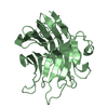
SHEET THE SHEET STRUCTURE OF THIS MOLECULE IS BIFURCATED. IN ORDER TO REPRESENT THIS FEATURE IN ... SHEET THE SHEET STRUCTURE OF THIS MOLECULE IS BIFURCATED. IN ORDER TO REPRESENT THIS FEATURE IN THE SHEET RECORDS BELOW, TWO SHEETS ARE DEFINED.
Mass: 18.015 Da / Num. of mol.: 204 / Source method: isolated from a natural source / Formula: H2O
Compound details
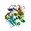
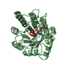
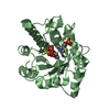
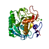
PRIA IS INVOLVED IN BOTH THE HISTIDINE AND TRYPTOPHAN BIOSYNTHETIC PATHWAYS. MEMBER OF THE ...PRIA IS INVOLVED IN BOTH THE HISTIDINE AND TRYPTOPHAN BIOSYNTHETIC PATHWAYS. MEMBER OF THE HISA/TRPF FAMILY OF PROTEINS.
-
Experimental details
-
Experiment
Experiment
Method: X-RAY DIFFRACTION / Number of used crystals: 1
-
Sample preparation
Crystal
Density Matthews: 2.3 Å3/Da / Density % sol: 47 % / Description: NONE
Crystal grow
pH: 4.8 Details: 1.50 M AMMONIUM SULFATE AND 100 MM SODIUM CITRATE PH 4.8
 Movie
Movie Controller


 Open data
 Basic information
 Components
 Keywords
 Function and homology information
 STREPTOMYCES COELICOLOR (bacteria)
 X-RAY DIFFRACTION /
  Authors
 Citation
 Structure visualization
 Downloads & links
 Other downloads
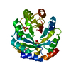


 PDBj
PDBj
 Assembly

 Mass: 96.063 Da / Num. of mol.: 2 / Source method: obtained synthetically / Formula: SO4
 Mass: 18.015 Da / Num. of mol.: 204 / Source method: isolated from a natural source / Formula: H2O
 Sample preparation
 / Beamline: ID14-1 / Wavelength: 0.934
  Processing