 Movie
Movie Controller

[English] 日本語
 Yorodumi
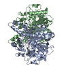
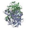
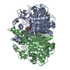
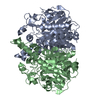
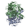
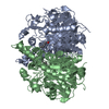
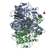
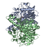
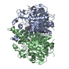
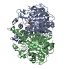
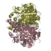
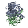
- PDB-2vba: beta-ketoacyl-ACP synthase I (KAS) from E. coli with bound amino-... -
+ Open data

- Basic information
| Entry | Database: PDB / ID: 2vba | ||||||
|---|---|---|---|---|---|---|---|
| Title | beta-ketoacyl-ACP synthase I (KAS) from E. coli with bound amino- thiazole inhibitor | ||||||
 Components Components | 3-OXOACYL-[ACYL-CARRIER-PROTEIN] SYNTHASE 1 | ||||||
 Keywords Keywords | TRANSFERASE / CYTOPLASM / ANTIBIOTIC / AMINO-THIAZOLE / ACYLTRANSFERASE / LIPID SYNTHESIS / FATTY ACID SYNTHESIS / FATTY ACID BIOSYNTHESIS | ||||||
| Function / homology |  Function and homology information Function and homology informationmonounsaturated fatty acid biosynthetic process / beta-ketoacyl-[acyl-carrier-protein] synthase I / 3-oxoacyl-[acyl-carrier-protein] synthase activity / fatty acid biosynthetic process / cytosol Similarity search - Function | ||||||
| Biological species |  | ||||||
| Method |  X-RAY DIFFRACTION / SYNCHROTRON / MOLECULAR REPLACEMENT / Resolution: 1.36 Å X-RAY DIFFRACTION / SYNCHROTRON / MOLECULAR REPLACEMENT / Resolution: 1.36 Å | ||||||
 Authors Authors | Pappenberger, G. / Schulz-Gasch, T. / Bailly, J. / Hennig, M. | ||||||
 Citation Citation | Journal: Acta Crystallogr.,Sect.D / Year: 2007 Title: Structure-Assisted Discovery of an Aminothiazole Derivative as a Lead Molecule for Inhibition of Bacterial Fatty-Acid Synthesis. Authors: Pappenberger, G. / Schulz-Gasch, T. / Kusznir, E. / Mueller, F. / Hennig, M. | ||||||
| History |
|
- Structure visualization
| Structure viewer | Molecule: MolmilJmol/JSmol |
|---|
- Downloads & links
-Download
| PDBx/mmCIF format | 2vba.cif.gz | 684.4 KB | Display | PDBx/mmCIF format |
|---|---|---|---|---|
| PDB format | pdb2vba.ent.gz | 573.6 KB | Display | PDB format |
| PDBx/mmJSON format | 2vba.json.gz | Tree view | PDBx/mmJSON format | |
| Others |  Other downloads Other downloads |
-Validation report
| Arichive directory | https://data.pdbj.org/pub/pdb/validation_reports/vb/2vbaftp://data.pdbj.org/pub/pdb/validation_reports/vb/2vba | HTTPS FTP |
|---|
-Related structure data
| Related structure data | 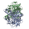 2vb7C 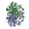 2vb8C 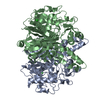 2vb9C  1ek4S S: Starting model for refinement C: citing same article ( |
|---|---|
| Similar structure data |


-Links
 PDBj
PDBj

- Assembly
| Deposited unit | 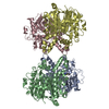
| ||||||||
|---|---|---|---|---|---|---|---|---|---|
| 1 | 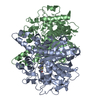
| ||||||||
| 2 | 
| ||||||||
| Unit cell |
|
-Components
| #1: Protein | Mass: 42656.203 Da / Num. of mol.: 4 Source method: isolated from a genetically manipulated source Source: (gene. exp.) References: UniProt: P0A953, beta-ketoacyl-[acyl-carrier-protein] synthase I #2: Chemical | ChemComp-P4T / |   Mass: 232.301 Da / Num. of mol.: 1 / Source method: obtained synthetically / Formula: C12H12N2OS Mass: 232.301 Da / Num. of mol.: 1 / Source method: obtained synthetically / Formula: C12H12N2OS#3: Water | ChemComp-HOH / |  Mass: 18.015 Da / Num. of mol.: 2431 / Source method: isolated from a natural source / Formula: H2O Mass: 18.015 Da / Num. of mol.: 2431 / Source method: isolated from a natural source / Formula: H2O |
|---|
-Experimental details
-Experiment
| Experiment | Method: X-RAY DIFFRACTION / Number of used crystals: 1 |
|---|
- Sample preparation
| Crystal | Density Matthews: 2.4 Å3/Da / Density % sol: 48 % / Description: NONE |
|---|---|
| Crystal grow | Method: vapor diffusion, hanging drop / pH: 7.5 Details: CRYSTALLIZATION CONDITIONS: HANGING DROP AGAINST 3% PEG 400, 1.9 M AMMONIUM SULPHATE, 0.1 M TRIS PH 7.5 SOAK WITH 0.01M LIGAND IN 30% PEG8000, 0.1M AMMONIUM SULPHATE, 0.1M TRIS PH7.5, 0.005M TCEP |
-Data collection
| Diffraction | Mean temperature: 100 K |
|---|---|
| Diffraction source | Source: SYNCHROTRON / Site: SLS  / Beamline: X06SA / Wavelength: 0.97881 / Beamline: X06SA / Wavelength: 0.97881 |
| Detector | Type: MARRESEARCH / Detector: CCD / Date: Sep 1, 2005 |
| Radiation | Protocol: SINGLE WAVELENGTH / Monochromatic (M) / Laue (L): M / Scattering type: x-ray |
| Radiation wavelength | Wavelength: 0.97881 Å / Relative weight: 1 |
| Reflection | Resolution: 1.36→20 Å / Num. obs: 685131 / % possible obs: 92.3 % / Observed criterion σ(I): 5 / Redundancy: 3 % / Rmerge(I) obs: 0.06 / Net I/σ(I): 13.5 |
| Reflection shell | Resolution: 1.35→1.45 Å / Redundancy: 2.7 % / Rmerge(I) obs: 0.2 / Mean I/σ(I) obs: 6.2 / % possible all: 72.9 |
- Processing
| Software |
| ||||||||||||||||||||||||||||||||||||||||||||||||||||||||||||||||||||||||||||||||||||||||||||||||||||||||||||||||||||||||||||||||||||||||||||||||||||||||||||||||||||||||||||||||||||||
|---|---|---|---|---|---|---|---|---|---|---|---|---|---|---|---|---|---|---|---|---|---|---|---|---|---|---|---|---|---|---|---|---|---|---|---|---|---|---|---|---|---|---|---|---|---|---|---|---|---|---|---|---|---|---|---|---|---|---|---|---|---|---|---|---|---|---|---|---|---|---|---|---|---|---|---|---|---|---|---|---|---|---|---|---|---|---|---|---|---|---|---|---|---|---|---|---|---|---|---|---|---|---|---|---|---|---|---|---|---|---|---|---|---|---|---|---|---|---|---|---|---|---|---|---|---|---|---|---|---|---|---|---|---|---|---|---|---|---|---|---|---|---|---|---|---|---|---|---|---|---|---|---|---|---|---|---|---|---|---|---|---|---|---|---|---|---|---|---|---|---|---|---|---|---|---|---|---|---|---|---|---|---|---|
| Refinement | Method to determine structure: MOLECULAR REPLACEMENT Starting model: PDB ENTRY 1EK4 Resolution: 1.36→19.87 Å / Cor.coef. Fo:Fc: 0.977 / Cor.coef. Fo:Fc free: 0.969 / SU B: 1.18 / SU ML: 0.022 / Cross valid method: THROUGHOUT / ESU R: 0.044 / ESU R Free: 0.044 / Stereochemistry target values: MAXIMUM LIKELIHOOD / Details: HYDROGENS HAVE BEEN ADDED IN THE RIDING POSITIONS.
| ||||||||||||||||||||||||||||||||||||||||||||||||||||||||||||||||||||||||||||||||||||||||||||||||||||||||||||||||||||||||||||||||||||||||||||||||||||||||||||||||||||||||||||||||||||||
| Solvent computation | Ion probe radii: 0.8 Å / Shrinkage radii: 0.8 Å / VDW probe radii: 1.2 Å / Solvent model: MASK | ||||||||||||||||||||||||||||||||||||||||||||||||||||||||||||||||||||||||||||||||||||||||||||||||||||||||||||||||||||||||||||||||||||||||||||||||||||||||||||||||||||||||||||||||||||||
| Displacement parameters | Biso mean: 17.49 Å2
| ||||||||||||||||||||||||||||||||||||||||||||||||||||||||||||||||||||||||||||||||||||||||||||||||||||||||||||||||||||||||||||||||||||||||||||||||||||||||||||||||||||||||||||||||||||||
| Refinement step | Cycle: LAST / Resolution: 1.36→19.87 Å
| ||||||||||||||||||||||||||||||||||||||||||||||||||||||||||||||||||||||||||||||||||||||||||||||||||||||||||||||||||||||||||||||||||||||||||||||||||||||||||||||||||||||||||||||||||||||
| Refine LS restraints |
|