 Movie
Movie Controller

[English] 日本語
 Yorodumi
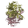
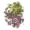
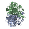
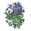
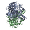
- PDB-1dd8: CRYSTAL STRUCTURE OF BETA-KETOACYL-[ACYL CARRIER PROTEIN] SYNTHAS... -
+ Open data

- Basic information
| Entry | Database: PDB / ID: 1dd8 | ||||||
|---|---|---|---|---|---|---|---|
| Title | CRYSTAL STRUCTURE OF BETA-KETOACYL-[ACYL CARRIER PROTEIN] SYNTHASE I FROM ESCHERICHIA COLI | ||||||
 Components Components | BETA-KETOACYL [ACYL CARRIER PROTEIN] SYNTHASE I | ||||||
 Keywords Keywords | TRANSFERASE / THIOLASE FOLD | ||||||
| Function / homology |  Function and homology information Function and homology informationmonounsaturated fatty acid biosynthetic process / beta-ketoacyl-[acyl-carrier-protein] synthase I / 3-oxoacyl-[acyl-carrier-protein] synthase activity / fatty acid biosynthetic process / cytosol Similarity search - Function | ||||||
| Biological species |  | ||||||
| Method |  X-RAY DIFFRACTION / Resolution: 2.3 Å X-RAY DIFFRACTION / Resolution: 2.3 Å | ||||||
 Authors Authors | Olsen, J.G. / Kadziola, A. / von Wettstein-Knowles, P. / Siggaard-Andersen, M. / Lindquist, Y. / Larsen, S. | ||||||
 Citation Citation | Journal: FEBS Lett. / Year: 1999 Title: The X-ray crystal structure of beta-ketoacyl [acyl carrier protein] synthase I. Authors: Olsen, J.G. / Kadziola, A. / von Wettstein-Knowles, P. / Siggaard-Andersen, M. / Lindquist, Y. / Larsen, S. | ||||||
| History |
|
- Structure visualization
| Structure viewer | Molecule: MolmilJmol/JSmol |
|---|
- Downloads & links
-Download
| PDBx/mmCIF format | 1dd8.cif.gz | 303.3 KB | Display | PDBx/mmCIF format |
|---|---|---|---|---|
| PDB format | pdb1dd8.ent.gz | 248.9 KB | Display | PDB format |
| PDBx/mmJSON format | 1dd8.json.gz | Tree view | PDBx/mmJSON format | |
| Others |  Other downloads Other downloads |
-Validation report
| Arichive directory | https://data.pdbj.org/pub/pdb/validation_reports/dd/1dd8ftp://data.pdbj.org/pub/pdb/validation_reports/dd/1dd8 | HTTPS FTP |
|---|
-Related structure data
| Similar structure data |
|---|


-Links
 PDBj
PDBj

- Assembly
| Deposited unit | 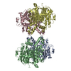
| ||||||||
|---|---|---|---|---|---|---|---|---|---|
| 1 | 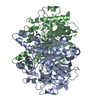
| ||||||||
| 2 | 
| ||||||||
| Unit cell |
|
-Components
| #1: Protein | Mass: 42684.254 Da / Num. of mol.: 4 / Source method: isolated from a natural source / Source: (natural) References: UniProt: P0A953, beta-ketoacyl-[acyl-carrier-protein] synthase I #2: Water | ChemComp-HOH / |  Mass: 18.015 Da / Num. of mol.: 508 / Source method: isolated from a natural source / Formula: H2O Mass: 18.015 Da / Num. of mol.: 508 / Source method: isolated from a natural source / Formula: H2O |
|---|
-Experimental details
-Experiment
| Experiment | Method: X-RAY DIFFRACTION / Number of used crystals: 1 |
|---|
- Sample preparation
| Crystal | Density Matthews: 2.66 Å3/Da / Density % sol: 53.73 % | ||||||||||||||||||||
|---|---|---|---|---|---|---|---|---|---|---|---|---|---|---|---|---|---|---|---|---|---|
| Crystal grow | Temperature: 293 K / Method: vapor diffusion, hanging drop / pH: 7.4 Details: bis-tris propane, ammonium sulfate, PEG400, pH 7.4, VAPOR DIFFUSION, HANGING DROP, temperature 20K | ||||||||||||||||||||
| Crystal grow | *PLUS pH: 7.5 / Details: Olsen, J.G., (1995) Protein Pept. Lett., 1, 246. | ||||||||||||||||||||
| Components of the solutions | *PLUS
|
-Data collection
| Diffraction | Mean temperature: 298 K |
|---|---|
| Diffraction source | Source: ROTATING ANODE / Type: RIGAKU RU200 / Wavelength: 1.5418 |
| Detector | Type: RIGAKU RAXIS II / Detector: IMAGE PLATE / Date: Jun 15, 1996 |
| Radiation | Protocol: SINGLE WAVELENGTH / Monochromatic (M) / Laue (L): M / Scattering type: x-ray |
| Radiation wavelength | Wavelength: 1.5418 Å / Relative weight: 1 |
| Reflection | Resolution: 2.3→30 Å / Num. all: 76863 / Num. obs: 76863 / % possible obs: 94.3 % / Observed criterion σ(F): 0 / Observed criterion σ(I): 0 / Redundancy: 3.24 % / Rmerge(I) obs: 0.071 |
| Reflection shell | Resolution: 2.3→30 Å / Redundancy: 1.44 % / Rmerge(I) obs: 0.119 / Num. unique all: 2025 / % possible all: 51 |
| Reflection | *PLUS |
| Reflection shell | *PLUS % possible obs: 51 % |
- Processing
| Software |
| |||||||||||||||||||||||||
|---|---|---|---|---|---|---|---|---|---|---|---|---|---|---|---|---|---|---|---|---|---|---|---|---|---|---|
| Refinement | Resolution: 2.3→10 Å / Stereochemistry target values: Engh & Huber / Details: NCS restraints were used throughout the refinement
| |||||||||||||||||||||||||
| Refinement step | Cycle: LAST / Resolution: 2.3→10 Å
| |||||||||||||||||||||||||
| Software | *PLUS Name: X-PLOR / Classification: refinement | |||||||||||||||||||||||||
| Refinement | *PLUS | |||||||||||||||||||||||||
| Solvent computation | *PLUS | |||||||||||||||||||||||||
| Displacement parameters | *PLUS Biso mean: 15 Å2 | |||||||||||||||||||||||||
| Refine LS restraints | *PLUS
|
