 Movie
Movie Controller

[English] 日本語
 Yorodumi
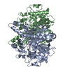
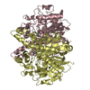
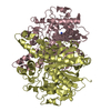
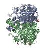
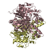
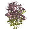
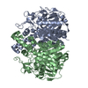
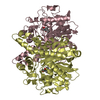
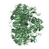
- PDB-1ek4: BETA-KETOACYL [ACYL CARRIER PROTEIN] SYNTHASE I IN COMPLEX WITH D... -
+ Open data

- Basic information
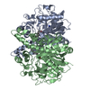
| Entry | Database: PDB / ID: 1ek4 | ||||||
|---|---|---|---|---|---|---|---|
| Title | BETA-KETOACYL [ACYL CARRIER PROTEIN] SYNTHASE I IN COMPLEX WITH DODECANOIC ACID TO 1.85 RESOLUTION | ||||||
 Components Components | BETA-KETOACYL [ACYL CARRIER PROTEIN] SYNTHASE I | ||||||
 Keywords Keywords | TRANSFERASE / fatty acid synthase / thiolase fold / Claisen condensation / fatty acid substrate complex | ||||||
| Function / homology |  Function and homology information Function and homology informationmonounsaturated fatty acid biosynthetic process / beta-ketoacyl-[acyl-carrier-protein] synthase I / 3-oxoacyl-[acyl-carrier-protein] synthase activity / fatty acid biosynthetic process / cytosol Similarity search - Function | ||||||
| Biological species |  | ||||||
| Method |  X-RAY DIFFRACTION / SYNCHROTRON / Resolution: 1.85 Å X-RAY DIFFRACTION / SYNCHROTRON / Resolution: 1.85 Å | ||||||
 Authors Authors | Olsen, J.G. / Kadziola, A. / Siggaard-Andersen, M. / von Wettstein-Knowles, P. / Larsen, S. | ||||||
 Citation Citation | Journal: Structure / Year: 2001 Title: Structures of beta-ketoacyl-acyl carrier protein synthase I complexed with fatty acids elucidate its catalytic machinery. Authors: Olsen, J.G. / Kadziola, A. / von Wettstein-Knowles, P. / Siggaard-Andersen, M. / Larsen, S. #1: Journal: FEBS Lett. / Year: 1999Title: The X-ray crystal structure of beta-ketoacyl [acyl carrier protein] synthase I Authors: Olsen, J.G. / Kadziola, A. / von Wettstein-Knowles, P. / Siggaard-Andersen, M. / Lindquist, Y. / Larsen, S. | ||||||
| History |
|
- Structure visualization
| Structure viewer | Molecule: MolmilJmol/JSmol |
|---|
- Downloads & links
-Download
| PDBx/mmCIF format | 1ek4.cif.gz | 314.3 KB | Display | PDBx/mmCIF format |
|---|---|---|---|---|
| PDB format | pdb1ek4.ent.gz | 255.1 KB | Display | PDB format |
| PDBx/mmJSON format | 1ek4.json.gz | Tree view | PDBx/mmJSON format | |
| Others |  Other downloads Other downloads |
-Validation report
| Arichive directory | https://data.pdbj.org/pub/pdb/validation_reports/ek/1ek4ftp://data.pdbj.org/pub/pdb/validation_reports/ek/1ek4 | HTTPS FTP |
|---|
-Related structure data


-Links
 PDBj
PDBj


- Assembly
| Deposited unit | 
| ||||||||
|---|---|---|---|---|---|---|---|---|---|
| 1 | 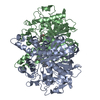
| ||||||||
| 2 | 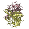
| ||||||||
| Unit cell |
| ||||||||
| Details | The biological entity is a dimer. There are two dimers in the asymmetric unit, one defined by chains A and B and one defined by chains C and D |
-Components
| #1: Protein | Mass: 44073.707 Da / Num. of mol.: 4 Mutation: C163S, MRGSHHHHHHGS N-TERMINAL HIS-TAG (NOT IN MODEL) Source method: isolated from a natural source / Source: (natural) References: UniProt: P0A953, beta-ketoacyl-[acyl-carrier-protein] synthase I #2: Chemical | ChemComp-DAO /   Mass: 200.318 Da / Num. of mol.: 4 / Source method: obtained synthetically / Formula: C12H24O2 Mass: 200.318 Da / Num. of mol.: 4 / Source method: obtained synthetically / Formula: C12H24O2#3: Water | ChemComp-HOH / |  Mass: 18.015 Da / Num. of mol.: 786 / Source method: isolated from a natural source / Formula: H2O Mass: 18.015 Da / Num. of mol.: 786 / Source method: isolated from a natural source / Formula: H2OHas protein modification | Y | |
|---|
-Experimental details
-Experiment
| Experiment | Method: X-RAY DIFFRACTION / Number of used crystals: 1 |
|---|
- Sample preparation
| Crystal | Density Matthews: 2.57 Å3/Da / Density % sol: 52.16 % | |||||||||||||||||||||||||
|---|---|---|---|---|---|---|---|---|---|---|---|---|---|---|---|---|---|---|---|---|---|---|---|---|---|---|
| Crystal grow | Temperature: 294 K / Method: vapor diffusion, hanging drop / pH: 7.4 Details: 1.8 M ammonium sulfate, 2 % PEG400, 0.1 M bis-Tris propane, pH 6.5, resulting pH 7.4, VAPOR DIFFUSION, HANGING DROP, temperature 294K | |||||||||||||||||||||||||
| Crystal grow | *PLUS | |||||||||||||||||||||||||
| Components of the solutions | *PLUS
|
-Data collection
| Diffraction | Mean temperature: 260 K |
|---|---|
| Diffraction source | Source: SYNCHROTRON / Site: MAX II  / Beamline: I711 / Wavelength: 1 / Beamline: I711 / Wavelength: 1 |
| Detector | Type: MARRESEARCH / Detector: AREA DETECTOR / Date: Nov 21, 1998 |
| Radiation | Protocol: SINGLE WAVELENGTH / Monochromatic (M) / Laue (L): M / Scattering type: x-ray |
| Radiation wavelength | Wavelength: 1 Å / Relative weight: 1 |
| Reflection | Resolution: 1.85→28.92 Å / Num. obs: 131071 / % possible obs: 85 % / Biso Wilson estimate: 10.1 Å2 / Rmerge(I) obs: 0.052 |
| Reflection shell | Resolution: 1.85→1.97 Å / Rmerge(I) obs: 0.267 / Num. unique all: 15875 / % possible all: 65.1 |
| Reflection | *PLUS % possible obs: 84 % / Rmerge(I) obs: 0.049 |
- Processing
| Software |
| ||||||||||||||||
|---|---|---|---|---|---|---|---|---|---|---|---|---|---|---|---|---|---|
| Refinement | Resolution: 1.85→28.92 Å / σ(F): 0 / σ(I): 0 / Stereochemistry target values: Eng & Huber
| ||||||||||||||||
| Refinement step | Cycle: LAST / Resolution: 1.85→28.92 Å
| ||||||||||||||||
| Refine LS restraints |
| ||||||||||||||||
| Software | *PLUS Name: CNS / Version: 0.9 / Classification: refinement | ||||||||||||||||
| Refine LS restraints | *PLUS
|
