 Movie
Movie Controller


+ Open data

- Basic information
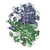
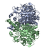
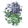
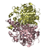
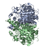
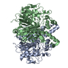
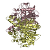
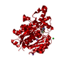
| Entry | Database: PDB / ID: 2buh | |||||||||
|---|---|---|---|---|---|---|---|---|---|---|
| Title | E. COLI BETA-KETOACYL (ACYL CARRIER PROTEIN) SYNTHASE I, 120 K | |||||||||
 Components Components | 3-OXOACYL-[ACYL-CARRIER-PROTEIN] SYNTHASE I | |||||||||
 Keywords Keywords | SYNTHASE / FATTY ACID SYNTHASE / THIOLASE FOLD / CLAISEN CONDENSATION / TRANSFERASE | |||||||||
| Function / homology |  Function and homology information Function and homology informationmonounsaturated fatty acid biosynthetic process / beta-ketoacyl-[acyl-carrier-protein] synthase I / 3-oxoacyl-[acyl-carrier-protein] synthase activity / fatty acid biosynthetic process / cytosol Similarity search - Function | |||||||||
| Biological species |  | |||||||||
| Method |  X-RAY DIFFRACTION / MOLECULAR REPLACEMENT / Resolution: 1.9 Å X-RAY DIFFRACTION / MOLECULAR REPLACEMENT / Resolution: 1.9 Å | |||||||||
 Authors Authors | Olsen, J.G. / Von Wettstein-Knowles, P. / Henriksen, A. | |||||||||
 Citation Citation | Journal: FEBS J. / Year: 2006 Title: Fatty acid synthesis. Role of active site histidines and lysine in Cys-His-His-type beta-ketoacyl-acyl carrier protein synthases. Authors: von Wettstein-Knowles, P. / Olsen, J.G. / McGuire, K.A. / Henriksen, A. | |||||||||
| History |
|
- Structure visualization
| Structure viewer | Molecule: MolmilJmol/JSmol |
|---|
- Downloads & links
-Download
| PDBx/mmCIF format | 2buh.cif.gz | 316.9 KB | Display | PDBx/mmCIF format |
|---|---|---|---|---|
| PDB format | pdb2buh.ent.gz | 258.8 KB | Display | PDB format |
| PDBx/mmJSON format | 2buh.json.gz | Tree view | PDBx/mmJSON format | |
| Others |  Other downloads Other downloads |
-Validation report
| Arichive directory | https://data.pdbj.org/pub/pdb/validation_reports/bu/2buhftp://data.pdbj.org/pub/pdb/validation_reports/bu/2buh | HTTPS FTP |
|---|
-Related structure data
| Related structure data | 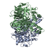 1h4fC 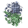 2buiC  2bywC  2byxC 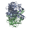 2byyC  2byzC 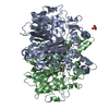 2bz3C  2bz4C 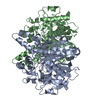 1dd8S S: Starting model for refinement C: citing same article ( |
|---|---|
| Similar structure data |


-Links
 PDBj
PDBj


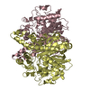
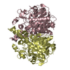
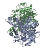
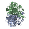
- Assembly
| Deposited unit | 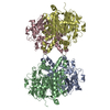
| ||||||||
|---|---|---|---|---|---|---|---|---|---|
| 1 | 
| ||||||||
| 2 | 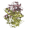
| ||||||||
| Unit cell |
|
-Components
| #1: Protein | Mass: 42656.203 Da / Num. of mol.: 4 Source method: isolated from a genetically manipulated source Source: (gene. exp.) References: UniProt: P14926, UniProt: P0A953*PLUS, beta-ketoacyl-[acyl-carrier-protein] synthase I #2: Chemical | ChemComp-NH4 /   Mass: 18.038 Da / Num. of mol.: 4 / Source method: obtained synthetically / Formula: H4N Mass: 18.038 Da / Num. of mol.: 4 / Source method: obtained synthetically / Formula: H4N#3: Water | ChemComp-HOH / |  Mass: 18.015 Da / Num. of mol.: 897 / Source method: isolated from a natural source / Formula: H2O Mass: 18.015 Da / Num. of mol.: 897 / Source method: isolated from a natural source / Formula: H2OCompound details | CATALYTIC ACTIVITY: ACYL-[ACYL-CARRIER PROTEIN] + MALONYL-[ACYL- CARRIER PROTEIN] = 3-OXOACYL-[ACYL- ...CATALYTIC ACTIVITY: ACYL-[ACYL-CARRIER PROTEIN] + MALONYL-[ACYL- CARRIER PROTEIN] = 3-OXOACYL-[ACYL-CARRIER PROTEIN] + CO(2) + [ACYL-CARRIER PROTEIN]. | |
|---|
-Experimental details
-Experiment
| Experiment | Method: X-RAY DIFFRACTION / Number of used crystals: 1 |
|---|
- Sample preparation
| Crystal | Density Matthews: 2.4 Å3/Da / Density % sol: 48 % |
|---|---|
| Crystal grow | pH: 7.4 Details: 2% PEG 400, 1.9 M AMMONIUM SULPHATE,0.1 M BIS-TRIS PROPANE PH 6.5 |
-Data collection
| Diffraction | Mean temperature: 120 K |
|---|---|
| Diffraction source | Source: ROTATING ANODE / Type: RIGAKU RU300 / Wavelength: 1.5418 |
| Detector | Type: RIGAKU IMAGE PLATE / Detector: IMAGE PLATE / Date: Dec 10, 2002 / Details: MIRRORS |
| Radiation | Protocol: SINGLE WAVELENGTH / Monochromatic (M) / Laue (L): M / Scattering type: x-ray |
| Radiation wavelength | Wavelength: 1.5418 Å / Relative weight: 1 |
| Reflection | Resolution: 1.9→29.7 Å / Num. obs: 134141 / % possible obs: 89.9 % / Observed criterion σ(I): 0 / Redundancy: 2.2 % / Biso Wilson estimate: 18.8 Å2 / Rmerge(I) obs: 0.13 / Net I/σ(I): 4.4 |
| Reflection shell | Resolution: 1.9→2.01 Å / Redundancy: 1.6 % / Rmerge(I) obs: 0.53 / Mean I/σ(I) obs: 2 / % possible all: 60.8 |
- Processing
| Software |
| ||||||||||||||||||||||||||||||||||||||||||||||||||||||||||||||||||||||||||||||||
|---|---|---|---|---|---|---|---|---|---|---|---|---|---|---|---|---|---|---|---|---|---|---|---|---|---|---|---|---|---|---|---|---|---|---|---|---|---|---|---|---|---|---|---|---|---|---|---|---|---|---|---|---|---|---|---|---|---|---|---|---|---|---|---|---|---|---|---|---|---|---|---|---|---|---|---|---|---|---|---|---|---|
| Refinement | Method to determine structure: MOLECULAR REPLACEMENT Starting model: PDB ENTRY 1DD8 Resolution: 1.9→29.73 Å / Rfactor Rfree error: 0.003 / Isotropic thermal model: RESTRAINED / Cross valid method: THROUGHOUT / σ(F): 0 / Stereochemistry target values: MLF
| ||||||||||||||||||||||||||||||||||||||||||||||||||||||||||||||||||||||||||||||||
| Solvent computation | Solvent model: FLAT MODEL / Bsol: 52.8952 Å2 / ksol: 0.384986 e/Å3 | ||||||||||||||||||||||||||||||||||||||||||||||||||||||||||||||||||||||||||||||||
| Displacement parameters | Biso mean: 25.7 Å2
| ||||||||||||||||||||||||||||||||||||||||||||||||||||||||||||||||||||||||||||||||
| Refine analyze |
| ||||||||||||||||||||||||||||||||||||||||||||||||||||||||||||||||||||||||||||||||
| Refinement step | Cycle: LAST / Resolution: 1.9→29.73 Å
| ||||||||||||||||||||||||||||||||||||||||||||||||||||||||||||||||||||||||||||||||
| Refine LS restraints |
| ||||||||||||||||||||||||||||||||||||||||||||||||||||||||||||||||||||||||||||||||
| LS refinement shell | Resolution: 1.9→2.02 Å / Rfactor Rfree error: 0.013 / Total num. of bins used: 6
| ||||||||||||||||||||||||||||||||||||||||||||||||||||||||||||||||||||||||||||||||
| Xplor file |
|
