SHEET THE SHEET STRUCTURE OF THIS MOLECULE IS BIFURCATED. IN ORDER TO REPRESENT THIS FEATURE IN ... SHEET THE SHEET STRUCTURE OF THIS MOLECULE IS BIFURCATED. IN ORDER TO REPRESENT THIS FEATURE IN THE SHEET RECORDS BELOW, TWO SHEETS ARE DEFINED.
Mass: 18.015 Da / Num. of mol.: 460 / Source method: isolated from a natural source / Formula: H2O
Compound details
ENGINEERED RESIDUE IN CHAIN A, GLU 138 TO ASP ENGINEERED RESIDUE IN CHAIN B, GLU 138 TO ASP ...ENGINEERED RESIDUE IN CHAIN A, GLU 138 TO ASP ENGINEERED RESIDUE IN CHAIN B, GLU 138 TO ASP ENGINEERED RESIDUE IN CHAIN C, GLU 138 TO ASP ENGINEERED RESIDUE IN CHAIN D, GLU 138 TO ASP ENGINEERED RESIDUE IN CHAIN E, GLU 138 TO ASP ENGINEERED RESIDUE IN CHAIN F, GLU 138 TO ASP ENGINEERED RESIDUE IN CHAIN G, GLU 138 TO ASP ENGINEERED RESIDUE IN CHAIN H, GLU 138 TO ASP ENGINEERED RESIDUE IN CHAIN I, GLU 138 TO ASP ENGINEERED RESIDUE IN CHAIN J, GLU 138 TO ASP ENGINEERED RESIDUE IN CHAIN K, GLU 138 TO ASP ENGINEERED RESIDUE IN CHAIN L, GLU 138 TO ASP
-
Experimental details
-
Experiment
Experiment
Method: X-RAY DIFFRACTION / Number of used crystals: 1
-
Sample preparation
Crystal
Density Matthews: 2.1 Å3/Da / Density % sol: 41 % / Description: NONE
Crystal grow
pH: 7.5 / Details: 27.5 %PEG 400, 50 MM MGSO4, 0.1 M HEPES, PH 7.5
-
Data collection
Diffraction
Mean temperature: 100 K
Diffraction source
Source: SYNCHROTRON / Site: MAX II / Beamline: I911-3 / Wavelength: 1
Resolution: 2.2→20 Å / Cor.coef. Fo:Fc: 0.939 / Cor.coef. Fo:Fc free: 0.918 / SU B: 6.073 / SU ML: 0.156 / Cross valid method: THROUGHOUT / ESU R: 0.373 / ESU R Free: 0.23 / Stereochemistry target values: MAXIMUM LIKELIHOOD / Details: HYDROGENS HAVE BEEN ADDED IN THE RIDING POSITIONS.
Rfactor
Num. reflection
% reflection
Selection details
Rfree
0.239
5341
5 %
RANDOM
Rwork
0.203
-
-
-
obs
0.205
101282
96.7 %
-
Solvent computation
Ion probe radii: 0.8 Å / Shrinkage radii: 0.8 Å / VDW probe radii: 1.2 Å / Solvent model: MASK
 Movie
Movie Controller

 Yorodumi
 Open data
 Basic information
 Components
 Keywords
 Function and homology information
 X-RAY DIFFRACTION /
  Authors
 Citation
 Structure visualization
 Downloads & links
 Other downloads


 PDBj
PDBj
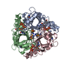
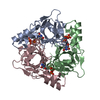
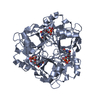
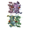
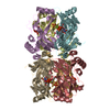
 Assembly
 Mass: 468.142 Da / Num. of mol.: 12 / Source method: obtained synthetically / Formula: C9H15N2O14P3
 Mass: 24.305 Da / Num. of mol.: 12 / Source method: obtained synthetically / Formula: Mg
 Mass: 96.063 Da / Num. of mol.: 4 / Source method: obtained synthetically / Formula: SO4
 Mass: 18.015 Da / Num. of mol.: 460 / Source method: isolated from a natural source / Formula: H2O
 Sample preparation
 / Beamline: I911-3 / Wavelength: 1
  Processing