 Movie
Movie Controller

[English] 日本語
 Yorodumi
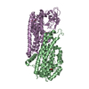
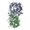
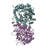
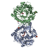
- PDB-3qom: Crystal structure of 6-phospho-beta-glucosidase from Lactobacillu... -
+ Open data

- Basic information
| Entry | Database: PDB / ID: 3qom | ||||||
|---|---|---|---|---|---|---|---|
| Title | Crystal structure of 6-phospho-beta-glucosidase from Lactobacillus plantarum | ||||||
 Components Components | 6-phospho-beta-glucosidase | ||||||
 Keywords Keywords | HYDROLASE / Structural Genomics / PSI-Biology / Midwest Center for Structural Genomics / MCSG / Glycoside hydrolase | ||||||
| Function / homology |  Function and homology information Function and homology information6-phospho-beta-glucosidase / 6-phospho-beta-glucosidase activity / carbohydrate catabolic process / cytosol Similarity search - Function | ||||||
| Biological species |  Lactobacillus plantarum (bacteria) Lactobacillus plantarum (bacteria) | ||||||
| Method |  X-RAY DIFFRACTION / SYNCHROTRON / SAD / Resolution: 1.498 Å X-RAY DIFFRACTION / SYNCHROTRON / SAD / Resolution: 1.498 Å | ||||||
 Authors Authors | Michalska, K. / Hatzos-Skintges, C. / Bearden, J. / Kohler, M. / Joachimiak, A. / Midwest Center for Structural Genomics (MCSG) | ||||||
 Citation Citation | Journal: Acta Crystallogr.,Sect.D / Year: 2013 Title: GH1-family 6-P-beta-glucosidases from human microbiome lactic acid bacteria. Authors: Michalska, K. / Tan, K. / Li, H. / Hatzos-Skintges, C. / Bearden, J. / Babnigg, G. / Joachimiak, A. | ||||||
| History |
|
- Structure visualization
| Structure viewer | Molecule: MolmilJmol/JSmol |
|---|
- Downloads & links
-Download
| PDBx/mmCIF format | 3qom.cif.gz | 219.6 KB | Display | PDBx/mmCIF format |
|---|---|---|---|---|
| PDB format | pdb3qom.ent.gz | 175.9 KB | Display | PDB format |
| PDBx/mmJSON format | 3qom.json.gz | Tree view | PDBx/mmJSON format | |
| Others |  Other downloads Other downloads |
-Validation report
| Arichive directory | https://data.pdbj.org/pub/pdb/validation_reports/qo/3qomftp://data.pdbj.org/pub/pdb/validation_reports/qo/3qom | HTTPS FTP |
|---|
-Related structure data
| Related structure data | 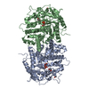 4f66C  4f79C 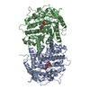 4gpnC  4gzeC C: citing same article ( |
|---|---|
| Similar structure data | |
| Other databases |


-Links
 PDBj
PDBj


- Assembly
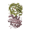
| Deposited unit | 
| ||||||||||||||||||||||||
|---|---|---|---|---|---|---|---|---|---|---|---|---|---|---|---|---|---|---|---|---|---|---|---|---|---|
| 1 | 
| ||||||||||||||||||||||||
| Unit cell |
| ||||||||||||||||||||||||
| Components on special symmetry positions |
| ||||||||||||||||||||||||
| Details | THE AUTHOR STATES THAT THE BIOLOGICAL UNIT OF THIS PROTEIN IS UNKNOWN. |
-Components
| #1: Protein | Mass: 55805.398 Da / Num. of mol.: 1 Source method: isolated from a genetically manipulated source Source: (gene. exp.) Lactobacillus plantarum (bacteria) / Strain: WCFS1 / Gene: pbg1, lp_0440 / Plasmid: pMCSG9 / Production host: References: UniProt: Q88ZA9, UniProt: F9UU25*PLUS, 6-phospho-beta-glucosidase | ||||||
|---|---|---|---|---|---|---|---|
| #2: Sugar | ChemComp-BGC /   Type: D-saccharide, beta linking / Mass: 180.156 Da / Num. of mol.: 1 Type: D-saccharide, beta linking / Mass: 180.156 Da / Num. of mol.: 1Source method: isolated from a genetically manipulated source Formula: C6H12O6 | ||||||
| #3: Chemical | ChemComp-PO4 /   Mass: 94.971 Da / Num. of mol.: 4 / Source method: obtained synthetically / Formula: PO4 Mass: 94.971 Da / Num. of mol.: 4 / Source method: obtained synthetically / Formula: PO4#4: Chemical | ChemComp-ACT / |   Mass: 59.044 Da / Num. of mol.: 1 / Source method: obtained synthetically / Formula: C2H3O2 Mass: 59.044 Da / Num. of mol.: 1 / Source method: obtained synthetically / Formula: C2H3O2#5: Water | ChemComp-HOH / |  Mass: 18.015 Da / Num. of mol.: 590 / Source method: isolated from a natural source / Formula: H2O Mass: 18.015 Da / Num. of mol.: 590 / Source method: isolated from a natural source / Formula: H2OHas protein modification | Y | |
-Experimental details
-Experiment
| Experiment | Method: X-RAY DIFFRACTION / Number of used crystals: 1 |
|---|
- Sample preparation
| Crystal | Density Matthews: 2.81 Å3/Da / Density % sol: 56.3 % |
|---|---|
| Crystal grow | Temperature: 297 K / Method: vapor diffusion, sitting drop / pH: 4.5 Details: 0.1 M sodium acetate, 0.8 M NaH2PO4/1.2 M K2HPO4, pH 4.5, VAPOR DIFFUSION, SITTING DROP, temperature 297K |
-Data collection
| Diffraction | Mean temperature: 100 K |
|---|---|
| Diffraction source | Source: SYNCHROTRON / Site: APS  / Beamline: 19-ID / Wavelength: 0.9791829 Å / Beamline: 19-ID / Wavelength: 0.9791829 Å |
| Detector | Type: ADSC QUANTUM 315r / Detector: CCD / Date: Dec 11, 2010 / Details: mirrors |
| Radiation | Monochromator: double crystal / Protocol: SINGLE WAVELENGTH / Monochromatic (M) / Laue (L): M / Scattering type: x-ray |
| Radiation wavelength | Wavelength: 0.9791829 Å / Relative weight: 1 |
| Reflection | Resolution: 1.5→50 Å / Num. all: 102436 / Num. obs: 102382 / % possible obs: 99.9 % / Observed criterion σ(I): -3 / Redundancy: 11.7 % / Biso Wilson estimate: 13.7 Å2 / Rmerge(I) obs: 0.112 / Net I/σ(I): 33.2 |
| Reflection shell | Resolution: 1.5→1.53 Å / Redundancy: 11.4 % / Rmerge(I) obs: 0.65 / Mean I/σ(I) obs: 3.7 / Num. unique all: 5046 / % possible all: 100 |
- Processing
| Software |
| ||||||||||||||||||||||||||||||||||||||||||||||||||||||||
|---|---|---|---|---|---|---|---|---|---|---|---|---|---|---|---|---|---|---|---|---|---|---|---|---|---|---|---|---|---|---|---|---|---|---|---|---|---|---|---|---|---|---|---|---|---|---|---|---|---|---|---|---|---|---|---|---|---|
| Refinement | Method to determine structure: SAD / Resolution: 1.498→31.048 Å / SU ML: 0.16 Isotropic thermal model: anisotropic for protein atoms, isotropic for solvent Cross valid method: THROUGHOUT / σ(F): 0 / Phase error: 10.59 / Stereochemistry target values: ML Details: HYDROGEN ATOMS HAVE BEEN ADDED IN THE RIDING POSITIONS
| ||||||||||||||||||||||||||||||||||||||||||||||||||||||||
| Solvent computation | Shrinkage radii: 0.9 Å / VDW probe radii: 1.11 Å / Solvent model: FLAT BULK SOLVENT MODEL / Bsol: 30.72 Å2 / ksol: 0.406 e/Å3 | ||||||||||||||||||||||||||||||||||||||||||||||||||||||||
| Displacement parameters |
| ||||||||||||||||||||||||||||||||||||||||||||||||||||||||
| Refinement step | Cycle: LAST / Resolution: 1.498→31.048 Å
| ||||||||||||||||||||||||||||||||||||||||||||||||||||||||
| Refine LS restraints |
| ||||||||||||||||||||||||||||||||||||||||||||||||||||||||
| LS refinement shell |
|
