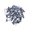
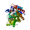
thioglucosidase / thioglucosidase activity / beta-glucosidase activity / carbohydrate metabolic process / metal ion binding Similarity search - Function
Glycosyl hydrolases family 1, N-terminal conserved site / Glycosyl hydrolases family 1 N-terminal signature. / Glycosyl hydrolase family 1 / Glycoside hydrolase family 1 / Glycosidases / Glycoside hydrolase superfamily / TIM Barrel / Alpha-Beta Barrel / Alpha Beta Similarity search - Domain/homology
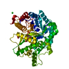
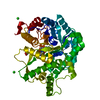
SHEET DETERMINATION METHOD: DSSP THE SHEETS PRESENTED AS "AA" IN EACH CHAIN ON SHEET RECORDS BELOW ... SHEET DETERMINATION METHOD: DSSP THE SHEETS PRESENTED AS "AA" IN EACH CHAIN ON SHEET RECORDS BELOW IS ACTUALLY AN 9-STRANDED BARREL THIS IS REPRESENTED BY A 10-STRANDED SHEET IN WHICH THE FIRST AND LAST STRANDS ARE IDENTICAL. THE SHEETS PRESENTED AS "BA" IN EACH CHAIN ON SHEET RECORDS BELOW IS ACTUALLY AN 8-STRANDED BARREL THIS IS REPRESENTED BY A 9-STRANDED SHEET IN WHICH THE FIRST AND LAST STRANDS ARE IDENTICAL.
Mass: 18.015 Da / Num. of mol.: 1450 / Source method: isolated from a natural source / Formula: H2O
Compound details
BELONGS TO FAMILY 1 OF GLYCOSYL HYDROLASES.
-
Experimental details
-
Experiment
Experiment
Method: X-RAY DIFFRACTION / Number of used crystals: 1
-
Sample preparation
Crystal
Density Matthews: 2.24 Å3/Da / Density % sol: 45.21 %
Crystal grow
Method: vapor diffusion, hanging drop / pH: 8 Details: APHID MYROSINASE IN 20 MM TRIS PH 8.0, 150 MM NACL, 10 MM MGCL2 AND 5MM DDT AT 10 MG/ML. CRYSTALS WERE GROWN IN HANGING DROPS WITH PRECIPITANT 0.2M SODIUM FORMATE, 20% PEG3350 MIXED TO THE ...Details: APHID MYROSINASE IN 20 MM TRIS PH 8.0, 150 MM NACL, 10 MM MGCL2 AND 5MM DDT AT 10 MG/ML. CRYSTALS WERE GROWN IN HANGING DROPS WITH PRECIPITANT 0.2M SODIUM FORMATE, 20% PEG3350 MIXED TO THE PROTEIN AT A RATIO OF 1:1.
Resolution: 1.1→19.65 Å / Cor.coef. Fo:Fc: 0.978 / Cor.coef. Fo:Fc free: 0.975 / SU B: 0.714 / SU ML: 0.016 / Cross valid method: THROUGHOUT / ESU R: 0.031 / ESU R Free: 0.03 / Stereochemistry target values: MAXIMUM LIKELIHOOD / Details: HYDROGENS HAVE BEEN ADDED IN THE RIDING POSITIONS.
Rfactor
Num. reflection
% reflection
Selection details
Rfree
0.146
16717
5.1 %
RANDOM
Rwork
0.132
-
-
-
obs
0.133
313715
85.9 %
-
Solvent computation
Ion probe radii: 0.8 Å / Shrinkage radii: 0.8 Å / VDW probe radii: 1.2 Å / Solvent model: MASK
 Movie
Movie Controller


 Open data
 Basic information
 Components
 Keywords
 Function and homology information
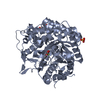
 BREVICORYNE BRASSICAE (cabbage aphid)
 X-RAY DIFFRACTION /
  Authors
 Citation
 Structure visualization
 Downloads & links
 Other downloads


 PDBj
PDBj Assembly
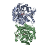


 Mass: 92.094 Da / Num. of mol.: 5 / Source method: obtained synthetically / Formula: C3H8O3
 Mass: 18.015 Da / Num. of mol.: 1450 / Source method: isolated from a natural source / Formula: H2O
 Sample preparation
 / Beamline: ID14-2 / Wavelength: 0.933
  Processing