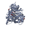
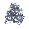
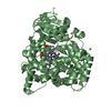
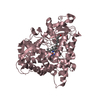
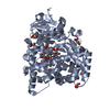
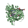
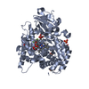
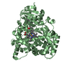
- PDB-2uwh: Cytochrome P450 BM3 mutant in complex with palmitic acid -
+
Open data
ID or keywords:
Loading...
-
Basic information
Entry
Database: PDB / ID: 2uwh
Title
Cytochrome P450 BM3 mutant in complex with palmitic acid
Components
BIFUNCTIONAL P-450\: NADPH-P450 REDUCTASE
Keywords
OXIDOREDUCTASE / FATTY-ACID BINDING / MULTIFUNCTIONAL ENZYME / METAL-BINDING / ELECTRON TRANSPORT / TRANSPORT / HYDROXYLASE / FLAVOPROTEIN / MONOOXYGENASE / FMN / FAD / NADP / IRON / HEME / REDOX / MEMBRANE / CYTOCHROME P450 BM3 MUTANT
Function / homology
Function and homology information
aromatase activity / NADPH-hemoprotein reductase / NADPH-hemoprotein reductase activity / oxidoreductase activity, acting on paired donors, with incorporation or reduction of molecular oxygen, reduced flavin or flavoprotein as one donor, and incorporation of one atom of oxygen / unspecific monooxygenase / FMN binding / flavin adenine dinucleotide binding / iron ion binding / heme binding / identical protein binding / cytosol Similarity search - Function
Mass: 18.015 Da / Num. of mol.: 519 / Source method: isolated from a natural source / Formula: H2O
Compound details
ENGINEERED RESIDUE IN CHAIN A, ALA 82 TO PHE ENGINEERED RESIDUE IN CHAIN B, ALA 82 TO PHE ...ENGINEERED RESIDUE IN CHAIN A, ALA 82 TO PHE ENGINEERED RESIDUE IN CHAIN B, ALA 82 TO PHE ENGINEERED RESIDUE IN CHAIN C, ALA 82 TO PHE ENGINEERED RESIDUE IN CHAIN D, ALA 82 TO PHE ENGINEERED RESIDUE IN CHAIN E, ALA 82 TO PHE ENGINEERED RESIDUE IN CHAIN F, ALA 82 TO PHE
-
Experimental details
-
Experiment
Experiment
Method: X-RAY DIFFRACTION / Number of used crystals: 1
-
Sample preparation
Crystal
Density Matthews: 2.44 Å3/Da / Density % sol: 49.26 % / Description: NONE
Crystal grow
pH: 5 Details: 140 MM MGCL2, 25% POLYETHYLENE GLYCOL 2000MME AND 100 MM MESA, PH 5.0
Resolution: 2.8→114.71 Å / Cor.coef. Fo:Fc: 0.915 / Cor.coef. Fo:Fc free: 0.84 / SU B: 18.329 / SU ML: 0.364 / Cross valid method: THROUGHOUT / ESU R: 1.527 / ESU R Free: 0.482 / Stereochemistry target values: MAXIMUM LIKELIHOOD / Details: HYDROGENS HAVE BEEN ADDED IN THE RIDING POSITIONS.
Rfactor
Num. reflection
% reflection
Selection details
Rfree
0.299
3858
5 %
RANDOM
Rwork
0.215
-
-
-
obs
0.219
72833
97.8 %
-
Solvent computation
Ion probe radii: 0.8 Å / Shrinkage radii: 0.8 Å / VDW probe radii: 1.4 Å / Solvent model: MASK
 Movie
Movie Controller


 Open data
 Basic information
 Components
 Keywords
 Function and homology information
 BACILLUS MEGATERIUM (bacteria)
 X-RAY DIFFRACTION /
  Authors
 Citation
 Structure visualization
 Downloads & links
 Other downloads


 PDBj
PDBj


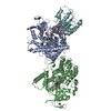
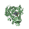
 Assembly


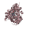
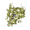


 Mass: 616.487 Da / Num. of mol.: 6 / Source method: obtained synthetically / Formula: C34H32FeN4O4
 Mass: 256.424 Da / Num. of mol.: 6 / Source method: obtained synthetically / Formula: C16H32O2
 Mass: 18.015 Da / Num. of mol.: 519 / Source method: isolated from a natural source / Formula: H2O
 Sample preparation
 / Beamline: ID14-3 / Wavelength: 0.931
  Processing