 Movie
Movie Controller


+ Open data

- Basic information
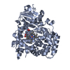
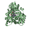
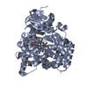
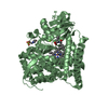
| Entry | Database: PDB / ID: 1fah | ||||||
|---|---|---|---|---|---|---|---|
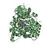
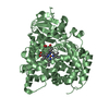
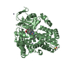
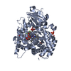
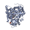
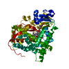
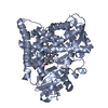
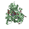
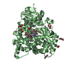
| Title | STRUCTURE OF CYTOCHROME P450 | ||||||
 Components Components | CYTOCHROME P450 BM-3 | ||||||
 Keywords Keywords | ELECTRON TRANSPORT / MONOOXYGENASE / HEME | ||||||
| Function / homology |  Function and homology information Function and homology informationaromatase activity / NADPH-hemoprotein reductase / NADPH-hemoprotein reductase activity / oxidoreductase activity, acting on paired donors, with incorporation or reduction of molecular oxygen, reduced flavin or flavoprotein as one donor, and incorporation of one atom of oxygen / unspecific monooxygenase / FMN binding / flavin adenine dinucleotide binding / iron ion binding / heme binding / identical protein binding / cytosol Similarity search - Function | ||||||
| Biological species |  Bacillus megaterium (bacteria) Bacillus megaterium (bacteria) | ||||||
| Method |  X-RAY DIFFRACTION / DIFFERENCE FOURIER / Resolution: 2.3 Å X-RAY DIFFRACTION / DIFFERENCE FOURIER / Resolution: 2.3 Å | ||||||
 Authors Authors | Li, H.Y. / Poulos, T.L. | ||||||
 Citation Citation | Journal: Biochemistry / Year: 1995 Title: The role of Thr268 in oxygen activation of cytochrome P450BM-3. Authors: Yeom, H. / Sligar, S.G. / Li, H. / Poulos, T.L. / Fulco, A.J. #1: Journal: Acta Crystallogr.,Sect.D / Year: 1995Title: Modeling Protein-Substrate Interactions in the Heme Domain of Cytochrome P450Bm-3 Authors: Li, H.Y. / Poulos, T.L. #2: Journal: Science / Year: 1993Title: Crystal Structure of Hemoprotein Domain of P450Bm-3, a Prototype for Microsomal P450'S Authors: Ravichandran, K.G. / Boddupalli, S.S. / Hasemann, C.A. / Peterson, J.A. / Deisenhofer, J. | ||||||
| History |
|
- Structure visualization
| Structure viewer | Molecule: MolmilJmol/JSmol |
|---|
- Downloads & links
-Download
| PDBx/mmCIF format | 1fah.cif.gz | 202.1 KB | Display | PDBx/mmCIF format |
|---|---|---|---|---|
| PDB format | pdb1fah.ent.gz | 162.1 KB | Display | PDB format |
| PDBx/mmJSON format | 1fah.json.gz | Tree view | PDBx/mmJSON format | |
| Others |  Other downloads Other downloads |
-Validation report
| Arichive directory | https://data.pdbj.org/pub/pdb/validation_reports/fa/1fahftp://data.pdbj.org/pub/pdb/validation_reports/fa/1fah | HTTPS FTP |
|---|
-Related structure data
| Similar structure data |
|---|


-Links
 PDBj
PDBj


- Assembly
| Deposited unit | 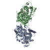
| ||||||||
|---|---|---|---|---|---|---|---|---|---|
| 1 | 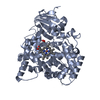
| ||||||||
| 2 | 
| ||||||||
| Unit cell |
| ||||||||
| Noncrystallographic symmetry (NCS) | NCS oper: (Code: given Matrix: (-0.999026, 0.019407, -0.039625), Vector: |
-Components
| #1: Protein | Mass: 53766.270 Da / Num. of mol.: 2 / Fragment: HEME DOMAIN / Mutation: T268A Source method: isolated from a genetically manipulated source Source: (gene. exp.) Bacillus megaterium (bacteria) / Strain: 14581 / Production host: #2: Chemical |   Mass: 616.487 Da / Num. of mol.: 2 / Source method: obtained synthetically / Formula: C34H32FeN4O4 Mass: 616.487 Da / Num. of mol.: 2 / Source method: obtained synthetically / Formula: C34H32FeN4O4#3: Water | ChemComp-HOH / |  Mass: 18.015 Da / Num. of mol.: 397 / Source method: isolated from a natural source / Formula: H2O Mass: 18.015 Da / Num. of mol.: 397 / Source method: isolated from a natural source / Formula: H2O |
|---|
-Experimental details
-Experiment
| Experiment | Method: X-RAY DIFFRACTION / Number of used crystals: 1 |
|---|
- Sample preparation
| Crystal | Density Matthews: 2.65 Å3/Da / Density % sol: 53 % | ||||||||||||||||||||||||||||||
|---|---|---|---|---|---|---|---|---|---|---|---|---|---|---|---|---|---|---|---|---|---|---|---|---|---|---|---|---|---|---|---|
| Crystal grow | pH: 6.8 / Details: SEE REFERENCE 1, pH 6.8 | ||||||||||||||||||||||||||||||
| Crystal grow | *PLUS Method: vapor diffusion, sitting drop | ||||||||||||||||||||||||||||||
| Components of the solutions | *PLUS
|
-Data collection
| Diffraction | Mean temperature: 296 K |
|---|---|
| Diffraction source | Source: ROTATING ANODE / Type: SIEMENS / Wavelength: 1.5418 |
| Detector | Type: SIEMENS-NICOLET X100 / Detector: AREA DETECTOR / Date: Sep 19, 1994 / Details: YES |
| Radiation | Monochromatic (M) / Laue (L): M / Scattering type: x-ray |
| Radiation wavelength | Wavelength: 1.5418 Å / Relative weight: 1 |
| Reflection | Resolution: 2.3→50 Å / Num. obs: 44522 / % possible obs: 88.2 % / Observed criterion σ(I): 0 / Redundancy: 3 % / Rmerge(I) obs: 0.08 / Rsym value: 0.08 / Net I/σ(I): 10.39 |
| Reflection shell | Resolution: 2.3→2.4 Å / Redundancy: 2 % / Rmerge(I) obs: 0.3 / Mean I/σ(I) obs: 1 / Rsym value: 0.3 / % possible all: 62.46 |
| Reflection | *PLUS Num. measured all: 120931 / Rmerge(I) obs: 0.08 |
| Reflection shell | *PLUS % possible obs: 62.5 % |
- Processing
| Software |
| ||||||||||||||||||||||||||||||||||||||||||||||||||||||||||||
|---|---|---|---|---|---|---|---|---|---|---|---|---|---|---|---|---|---|---|---|---|---|---|---|---|---|---|---|---|---|---|---|---|---|---|---|---|---|---|---|---|---|---|---|---|---|---|---|---|---|---|---|---|---|---|---|---|---|---|---|---|---|
| Refinement | Method to determine structure: DIFFERENCE FOURIER Starting model: WILD TYPE P450BM-3 HEME DOMAIN Resolution: 2.3→10 Å / σ(F): 2 Details: SIMILAR TO THE CRYSTAL STRUCTURE OF THE WILD TYPE P450BM-3 HEME DOMAIN, THE LOOP REGION BETWEEN THE F AND G HELICES IS POORLY DEFINED IN THE ELECTRON DENSITY AND THEREFORE MODELED WITH HIGH B FACTORS.
| ||||||||||||||||||||||||||||||||||||||||||||||||||||||||||||
| Refine analyze | Luzzati coordinate error obs: 0.25 Å | ||||||||||||||||||||||||||||||||||||||||||||||||||||||||||||
| Refinement step | Cycle: LAST / Resolution: 2.3→10 Å
| ||||||||||||||||||||||||||||||||||||||||||||||||||||||||||||
| Refine LS restraints |
| ||||||||||||||||||||||||||||||||||||||||||||||||||||||||||||
| Xplor file | Serial no: 1 / Param file: PARAM19X.HEME / Topol file: TOPH19X.HEME | ||||||||||||||||||||||||||||||||||||||||||||||||||||||||||||
| Software | *PLUS Name: X-PLOR / Version: 3.1 / Classification: refinement | ||||||||||||||||||||||||||||||||||||||||||||||||||||||||||||
| Refine LS restraints | *PLUS
|
