 Movie
Movie Controller


+ Open data

- Basic information
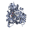
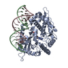
| Entry | Database: PDB / ID: 1fag | ||||||
|---|---|---|---|---|---|---|---|
| Title | STRUCTURE OF CYTOCHROME P450 | ||||||
 Components Components | CYTOCHROME P450 BM-3 | ||||||
 Keywords Keywords | ELECTRON TRANSPORT / MONOOXYGENASE / HEME | ||||||
| Function / homology |  Function and homology information Function and homology informationaromatase activity / NADPH-hemoprotein reductase / NADPH-hemoprotein reductase activity / oxidoreductase activity, acting on paired donors, with incorporation or reduction of molecular oxygen, reduced flavin or flavoprotein as one donor, and incorporation of one atom of oxygen / unspecific monooxygenase / FMN binding / flavin adenine dinucleotide binding / iron ion binding / heme binding / identical protein binding / cytosol Similarity search - Function | ||||||
| Biological species |  Bacillus megaterium (bacteria) Bacillus megaterium (bacteria) | ||||||
| Method |  X-RAY DIFFRACTION / SYNCHROTRON / MOLECULAR REPLACEMENT / Resolution: 2.7 Å X-RAY DIFFRACTION / SYNCHROTRON / MOLECULAR REPLACEMENT / Resolution: 2.7 Å | ||||||
 Authors Authors | Li, H.Y. / Poulos, T.L. | ||||||
 Citation Citation | Journal: Nat.Struct.Biol. / Year: 1997 Title: The structure of the cytochrome p450BM-3 haem domain complexed with the fatty acid substrate, palmitoleic acid. Authors: Li, H. / Poulos, T.L. #1: Journal: To be PublishedTitle: Comformational Dynamics in Cytochrome P450-Substrate Interactions Authors: Li, H.Y. / Poulos, T.L. #2: Journal: Acta Crystallogr.,Sect.D / Year: 1995Title: Modeling Protein-Substrate Interactions in the Heme Domain of Cytochrome P450Bm-3 Authors: Li, H.Y. / Poulos, T.L. #3: Journal: Science / Year: 1993Title: Crystal Structure of Hemoprotein Domain of P450Bm-3, a Prototype for Microsomal P450'S Authors: Ravichandran, K.G. / Boddupalli, S.S. / Hasemann, C.A. / Peterson, J.A. / Deisenhofer, J. | ||||||
| History |
|
- Structure visualization
| Structure viewer | Molecule: MolmilJmol/JSmol |
|---|
- Downloads & links
-Download
| PDBx/mmCIF format | 1fag.cif.gz | 369.9 KB | Display | PDBx/mmCIF format |
|---|---|---|---|---|
| PDB format | pdb1fag.ent.gz | 306.2 KB | Display | PDB format |
| PDBx/mmJSON format | 1fag.json.gz | Tree view | PDBx/mmJSON format | |
| Others |  Other downloads Other downloads |
-Validation report
| Arichive directory | https://data.pdbj.org/pub/pdb/validation_reports/fa/1fagftp://data.pdbj.org/pub/pdb/validation_reports/fa/1fag | HTTPS FTP |
|---|
-Related structure data
| Similar structure data |
|---|


-Links
 PDBj
PDBj


- Assembly
| Deposited unit | 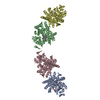
| ||||||||||||||||
|---|---|---|---|---|---|---|---|---|---|---|---|---|---|---|---|---|---|
| 1 | 
| ||||||||||||||||
| 2 | 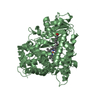
| ||||||||||||||||
| 3 | 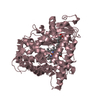
| ||||||||||||||||
| 4 | 
| ||||||||||||||||
| Unit cell |
| ||||||||||||||||
| Noncrystallographic symmetry (NCS) | NCS oper:
|
-Components
| #1: Protein | Mass: 53796.293 Da / Num. of mol.: 4 / Fragment: HEME DOMAIN Source method: isolated from a genetically manipulated source Source: (gene. exp.) Bacillus megaterium (bacteria) / Strain: 14581 / Production host: #2: Chemical | ChemComp-HEM /   Mass: 616.487 Da / Num. of mol.: 4 / Source method: obtained synthetically / Formula: C34H32FeN4O4 Mass: 616.487 Da / Num. of mol.: 4 / Source method: obtained synthetically / Formula: C34H32FeN4O4#3: Chemical | ChemComp-PAM / 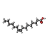  Mass: 254.408 Da / Num. of mol.: 4 / Source method: obtained synthetically / Formula: C16H30O2 Mass: 254.408 Da / Num. of mol.: 4 / Source method: obtained synthetically / Formula: C16H30O2 |
|---|
-Experimental details
-Experiment
| Experiment | Method: X-RAY DIFFRACTION / Number of used crystals: 1 |
|---|
- Sample preparation
| Crystal | Density Matthews: 2.28 Å3/Da / Density % sol: 46 % | ||||||||||||||||||||||||||||||||||||||||||||||||||||||
|---|---|---|---|---|---|---|---|---|---|---|---|---|---|---|---|---|---|---|---|---|---|---|---|---|---|---|---|---|---|---|---|---|---|---|---|---|---|---|---|---|---|---|---|---|---|---|---|---|---|---|---|---|---|---|---|
| Crystal grow | pH: 6 / Details: SEE REFERENCE 1, pH 6.0 | ||||||||||||||||||||||||||||||||||||||||||||||||||||||
| Crystal grow | *PLUS Method: vapor diffusion, sitting drop | ||||||||||||||||||||||||||||||||||||||||||||||||||||||
| Components of the solutions | *PLUS
|
-Data collection
| Diffraction | Mean temperature: 100 K |
|---|---|
| Diffraction source | Source: SYNCHROTRON / Site: SSRL  / Beamline: BL7-1 / Wavelength: 1.008 / Beamline: BL7-1 / Wavelength: 1.008 |
| Detector | Type: MARRESEARCH / Detector: IMAGE PLATE / Date: May 16, 1996 |
| Radiation | Monochromatic (M) / Laue (L): M / Scattering type: x-ray |
| Radiation wavelength | Wavelength: 1.008 Å / Relative weight: 1 |
| Reflection | Resolution: 2.6→48.8 Å / Num. obs: 60630 / % possible obs: 99 % / Observed criterion σ(I): 0 / Redundancy: 7 % / Rmerge(I) obs: 0.071 / Rsym value: 0.071 / Net I/σ(I): 8.3 |
| Reflection shell | Resolution: 2.7→2.8 Å / Redundancy: 7.2 % / Rmerge(I) obs: 0.5 / Mean I/σ(I) obs: 1.4 / Rsym value: 0.5 / % possible all: 97 |
- Processing
| Software |
| ||||||||||||||||||||||||||||||||||||||||||||||||||||||||||||
|---|---|---|---|---|---|---|---|---|---|---|---|---|---|---|---|---|---|---|---|---|---|---|---|---|---|---|---|---|---|---|---|---|---|---|---|---|---|---|---|---|---|---|---|---|---|---|---|---|---|---|---|---|---|---|---|---|---|---|---|---|---|
| Refinement | Method to determine structure: MOLECULAR REPLACEMENT Starting model: SUBSTRATE-FREE P450BM-3 HEME DOMAIN Resolution: 2.7→10 Å / σ(F): 2 Details: THE F/G AND G/H LOOP REGIONS IN FOUR MOLECULES SHOWED QUITE DIFFERENT CONFORMATIONS. A FEW RESIDUES IN THESE SURFACE LOOP REGIONS ARE NOT WELL DEFINED IN THE STRUCTURE WITH RATHER HIGH B ...Details: THE F/G AND G/H LOOP REGIONS IN FOUR MOLECULES SHOWED QUITE DIFFERENT CONFORMATIONS. A FEW RESIDUES IN THESE SURFACE LOOP REGIONS ARE NOT WELL DEFINED IN THE STRUCTURE WITH RATHER HIGH B FACTORS, E.G. GLU 228 IN MOLECULES A AND C, GLN 229 IN MOLECULE D, ASP 195 IN MOLECULE C. THE PHI, PSI TORTION ANGLES OF LEUCINE 437 IN ALL FOUR MOLECULES FALL IN THE DISALLOWED REGION, BUT THE ODD CONFORMER MIGHT RESULT FROM THE NON-BONDING INTERACTION BETWEEN THE SUBSTRATE AND ENZYME.
| ||||||||||||||||||||||||||||||||||||||||||||||||||||||||||||
| Refine analyze | Luzzati coordinate error obs: 0.4 Å | ||||||||||||||||||||||||||||||||||||||||||||||||||||||||||||
| Refinement step | Cycle: LAST / Resolution: 2.7→10 Å
| ||||||||||||||||||||||||||||||||||||||||||||||||||||||||||||
| Refine LS restraints |
| ||||||||||||||||||||||||||||||||||||||||||||||||||||||||||||
| Software | *PLUS Name: X-PLOR / Version: 3.1 / Classification: refinement | ||||||||||||||||||||||||||||||||||||||||||||||||||||||||||||
| Refine LS restraints | *PLUS
|
