 Movie
Movie Controller

[English] 日本語
 Yorodumi
- PDB-6k24: Structure of the Rhodium Mesoporphyrin IX-Reconstituted CYP102A1 ... -
+ Open data

- Basic information
| Entry | Database: PDB / ID: 6k24 | ||||||
|---|---|---|---|---|---|---|---|
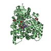
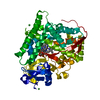
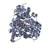
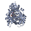
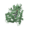
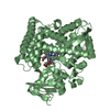
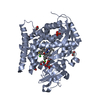
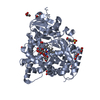
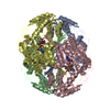
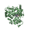
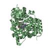
| Title | Structure of the Rhodium Mesoporphyrin IX-Reconstituted CYP102A1 Haem Domain with N-Abietoyl-L-Tryptophan | ||||||
 Components Components | Bifunctional cytochrome P450/NADPH--P450 reductase | ||||||
 Keywords Keywords | OXIDOREDUCTASE / Monooxygenase | ||||||
| Function / homology |  Function and homology information Function and homology informationaromatase activity / NADPH-hemoprotein reductase / NADPH-hemoprotein reductase activity / oxidoreductase activity, acting on paired donors, with incorporation or reduction of molecular oxygen, reduced flavin or flavoprotein as one donor, and incorporation of one atom of oxygen / unspecific monooxygenase / FMN binding / flavin adenine dinucleotide binding / iron ion binding / heme binding / identical protein binding / cytosol Similarity search - Function | ||||||
| Biological species |  Bacillus megaterium (bacteria) Bacillus megaterium (bacteria) | ||||||
| Method |  X-RAY DIFFRACTION / SYNCHROTRON / MOLECULAR REPLACEMENT / molecular replacement / Resolution: 2.1 Å X-RAY DIFFRACTION / SYNCHROTRON / MOLECULAR REPLACEMENT / molecular replacement / Resolution: 2.1 Å | ||||||
 Authors Authors | Stanfield, J.K. / Matsumoto, A. / Kasai, C. / Sugimoto, H. / Shiro, Y. / Watanabe, Y. / Shoji, O. | ||||||
| Funding support |  Japan, 1items Japan, 1items
| ||||||
 Citation Citation | Journal: Angew.Chem.Int.Ed.Engl. / Year: 2020 Title: Crystals in Minutes: Instant On-Site Microcrystallisation of Various Flavours of the CYP102A1 (P450BM3) Haem Domain. Authors: Stanfield, J.K. / Omura, K. / Matsumoto, A. / Kasai, C. / Sugimoto, H. / Shiro, Y. / Watanabe, Y. / Shoji, O. | ||||||
| History |
|
- Structure visualization
| Structure viewer | Molecule: MolmilJmol/JSmol |
|---|
- Downloads & links
-Download
| PDBx/mmCIF format | 6k24.cif.gz | 211.2 KB | Display | PDBx/mmCIF format |
|---|---|---|---|---|
| PDB format | pdb6k24.ent.gz | 166.5 KB | Display | PDB format |
| PDBx/mmJSON format | 6k24.json.gz | Tree view | PDBx/mmJSON format | |
| Others |  Other downloads Other downloads |
-Validation report
| Arichive directory | https://data.pdbj.org/pub/pdb/validation_reports/k2/6k24ftp://data.pdbj.org/pub/pdb/validation_reports/k2/6k24 | HTTPS FTP |
|---|
-Related structure data
| Related structure data |  6jlvC  6jmhC  6jmwC  6jo1C  6js8C  6jvcC  6jzsC  6k58C  6k9sC  3wspS S: Starting model for refinement C: citing same article ( |
|---|---|
| Similar structure data |


-Links
 PDBj
PDBj


- Assembly
| Deposited unit | 
| ||||||||
|---|---|---|---|---|---|---|---|---|---|
| 1 | 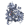
| ||||||||
| 2 | 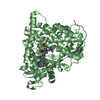
| ||||||||
| Unit cell |
|
-Components
| #1: Protein | Mass: 54979.508 Da / Num. of mol.: 2 Source method: isolated from a genetically manipulated source Source: (gene. exp.) Bacillus megaterium (bacteria) / Gene: cyp102A1 / Production host: References: UniProt: F2Q7T0, UniProt: P14779*PLUS, unspecific monooxygenase, NADPH-hemoprotein reductase #2: Chemical |   Mass: 488.661 Da / Num. of mol.: 2 / Source method: obtained synthetically / Formula: C31H40N2O3 Mass: 488.661 Da / Num. of mol.: 2 / Source method: obtained synthetically / Formula: C31H40N2O3#3: Chemical |   Mass: 667.580 Da / Num. of mol.: 2 / Source method: obtained synthetically / Formula: C34H36N4O4Rh Mass: 667.580 Da / Num. of mol.: 2 / Source method: obtained synthetically / Formula: C34H36N4O4Rh#4: Chemical |   Mass: 78.133 Da / Num. of mol.: 2 / Source method: obtained synthetically / Formula: C2H6OS / Comment: DMSO, precipitant*YM Mass: 78.133 Da / Num. of mol.: 2 / Source method: obtained synthetically / Formula: C2H6OS / Comment: DMSO, precipitant*YM#5: Water | ChemComp-HOH / |  Mass: 18.015 Da / Num. of mol.: 298 / Source method: isolated from a natural source / Formula: H2O Mass: 18.015 Da / Num. of mol.: 298 / Source method: isolated from a natural source / Formula: H2O |
|---|
-Experimental details
-Experiment
| Experiment | Method: X-RAY DIFFRACTION / Number of used crystals: 1 |
|---|
- Sample preparation
| Crystal | Density Matthews: 2.72 Å3/Da / Density % sol: 54.74 % |
|---|---|
| Crystal grow | Temperature: 293 K / Method: batch mode Details: PEG8000, Magnesium Chloride, Tris-HCl, 0.5% DMSO, 125uM N-Abietoyl-L-Tryptophan |
-Data collection
| Diffraction | Mean temperature: 100 K / Serial crystal experiment: N | ||||||||||||||||||||||||
|---|---|---|---|---|---|---|---|---|---|---|---|---|---|---|---|---|---|---|---|---|---|---|---|---|---|
| Diffraction source | Source: SYNCHROTRON / Site: SPring-8 / Beamline: BL26B1 / Wavelength: 1 Å | ||||||||||||||||||||||||
| Detector | Type: DECTRIS EIGER X 4M / Detector: PIXEL / Date: Oct 11, 2018 | ||||||||||||||||||||||||
| Radiation | Protocol: SINGLE WAVELENGTH / Monochromatic (M) / Laue (L): M / Scattering type: x-ray | ||||||||||||||||||||||||
| Radiation wavelength | Wavelength: 1 Å / Relative weight: 1 | ||||||||||||||||||||||||
| Reflection | Resolution: 2.1→48.74 Å / Num. obs: 60178 / % possible obs: 92.9 % / Redundancy: 6.7 % / CC1/2: 0.993 / Rmerge(I) obs: 0.103 / Rpim(I) all: 0.043 / Rrim(I) all: 0.112 / Net I/σ(I): 10.9 | ||||||||||||||||||||||||
| Reflection shell | Diffraction-ID: 1 / % possible all: 99.2
|
-Phasing
| Phasing | Method: molecular replacement |
|---|
- Processing
| Software |
| ||||||||||||||||||||||||||||||||||||||||||||||||||||||||||||
|---|---|---|---|---|---|---|---|---|---|---|---|---|---|---|---|---|---|---|---|---|---|---|---|---|---|---|---|---|---|---|---|---|---|---|---|---|---|---|---|---|---|---|---|---|---|---|---|---|---|---|---|---|---|---|---|---|---|---|---|---|---|
| Refinement | Method to determine structure: MOLECULAR REPLACEMENT Starting model: 3WSP Resolution: 2.1→48.74 Å / Cor.coef. Fo:Fc: 0.944 / Cor.coef. Fo:Fc free: 0.918 / SU B: 7.555 / SU ML: 0.186 / Cross valid method: THROUGHOUT / σ(F): 0 / ESU R: 0.258 / ESU R Free: 0.21 Details: HYDROGENS HAVE BEEN ADDED IN THE RIDING POSITIONS U VALUES : REFINED INDIVIDUALLY
| ||||||||||||||||||||||||||||||||||||||||||||||||||||||||||||
| Solvent computation | Ion probe radii: 0.8 Å / Shrinkage radii: 0.8 Å / VDW probe radii: 1.2 Å | ||||||||||||||||||||||||||||||||||||||||||||||||||||||||||||
| Displacement parameters | Biso max: 137.15 Å2 / Biso mean: 43.929 Å2 / Biso min: 21.74 Å2
| ||||||||||||||||||||||||||||||||||||||||||||||||||||||||||||
| Refinement step | Cycle: final / Resolution: 2.1→48.74 Å
| ||||||||||||||||||||||||||||||||||||||||||||||||||||||||||||
| Refine LS restraints |
| ||||||||||||||||||||||||||||||||||||||||||||||||||||||||||||
| LS refinement shell | Resolution: 2.1→2.154 Å / Rfactor Rfree error: 0 / Total num. of bins used: 20
|
