 Movie
Movie Controller

[English] 日本語
 Yorodumi
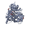
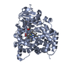
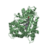
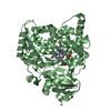
- PDB-1smj: Structure of the A264E mutant of cytochrome P450 BM3 complexed wi... -
+ Open data

- Basic information
| Entry | Database: PDB / ID: 1smj | ||||||
|---|---|---|---|---|---|---|---|
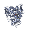
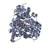
| Title | Structure of the A264E mutant of cytochrome P450 BM3 complexed with palmitoleate | ||||||
 Components Components | Bifunctional P-450:NADPH-P450 reductase | ||||||
 Keywords Keywords | OXIDOREDUCTASE / monooxygenase / fatty acid oxygenase / cytochrome P450 / substrate binding / palmitoleate | ||||||
| Function / homology |  Function and homology information Function and homology informationaromatase activity / NADPH-hemoprotein reductase / NADPH-hemoprotein reductase activity / oxidoreductase activity, acting on paired donors, with incorporation or reduction of molecular oxygen, reduced flavin or flavoprotein as one donor, and incorporation of one atom of oxygen / unspecific monooxygenase / FMN binding / flavin adenine dinucleotide binding / iron ion binding / heme binding / identical protein binding / cytosol Similarity search - Function | ||||||
| Biological species |  Bacillus megaterium (bacteria) Bacillus megaterium (bacteria) | ||||||
| Method |  X-RAY DIFFRACTION / SYNCHROTRON / MOLECULAR REPLACEMENT / Resolution: 2.75 Å X-RAY DIFFRACTION / SYNCHROTRON / MOLECULAR REPLACEMENT / Resolution: 2.75 Å | ||||||
 Authors Authors | Joyce, M.G. / Girvan, H.M. / Munro, A.W. / Leys, D. | ||||||
 Citation Citation | Journal: J.Biol.Chem. / Year: 2004 Title: A Single Mutation in Cytochrome P450 BM3 Induces the Conformational Rearrangement Seen upon Substrate Binding in the Wild-type Enzyme Authors: Joyce, M.G. / Girvan, H.M. / Munro, A.W. / Leys, D. | ||||||
| History |
|
- Structure visualization
| Structure viewer | Molecule: MolmilJmol/JSmol |
|---|
- Downloads & links
-Download
| PDBx/mmCIF format | 1smj.cif.gz | 375 KB | Display | PDBx/mmCIF format |
|---|---|---|---|---|
| PDB format | pdb1smj.ent.gz | 306.5 KB | Display | PDB format |
| PDBx/mmJSON format | 1smj.json.gz | Tree view | PDBx/mmJSON format | |
| Others |  Other downloads Other downloads |
-Validation report
| Arichive directory | https://data.pdbj.org/pub/pdb/validation_reports/sm/1smjftp://data.pdbj.org/pub/pdb/validation_reports/sm/1smj | HTTPS FTP |
|---|
-Related structure data
| Related structure data |  1smiC  1fagS S: Starting model for refinement C: citing same article ( |
|---|---|
| Similar structure data |


-Links
 PDBj
PDBj


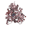
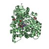
- Assembly
| Deposited unit | 
| ||||||||||
|---|---|---|---|---|---|---|---|---|---|---|---|
| 1 | 
| ||||||||||
| 2 | 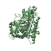
| ||||||||||
| 3 | 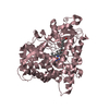
| ||||||||||
| 4 | 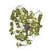
| ||||||||||
| Unit cell |
|
-Components
| #1: Protein | Mass: 53854.328 Da / Num. of mol.: 4 / Fragment: cytochrome P450 102 / Mutation: A264E Source method: isolated from a genetically manipulated source Source: (gene. exp.) Bacillus megaterium (bacteria) / Production host: #2: Chemical | ChemComp-HEM /   Mass: 616.487 Da / Num. of mol.: 4 / Source method: obtained synthetically / Formula: C34H32FeN4O4 Mass: 616.487 Da / Num. of mol.: 4 / Source method: obtained synthetically / Formula: C34H32FeN4O4#3: Chemical | ChemComp-PAM / 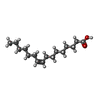  Mass: 254.408 Da / Num. of mol.: 4 / Source method: obtained synthetically / Formula: C16H30O2 Mass: 254.408 Da / Num. of mol.: 4 / Source method: obtained synthetically / Formula: C16H30O2Has protein modification | N | |
|---|
-Experimental details
-Experiment
| Experiment | Method: X-RAY DIFFRACTION / Number of used crystals: 1 |
|---|
- Sample preparation
| Crystal | Density Matthews: 2.41 Å3/Da / Density % sol: 48.6 % |
|---|---|
| Crystal grow | Temperature: 277 K / Method: vapor diffusion, sitting drop / pH: 6.3 Details: PEG 2000, MME, 100mM magnesium acetate, pH 6.3, VAPOR DIFFUSION, SITTING DROP, temperature 277K |
-Data collection
| Diffraction | Mean temperature: 100 K |
|---|---|
| Diffraction source | Source: SYNCHROTRON / Site: ESRF  / Beamline: ID14-1 / Wavelength: 0.9 Å / Beamline: ID14-1 / Wavelength: 0.9 Å |
| Detector | Type: ADSC QUANTUM 4 / Detector: CCD / Date: Aug 15, 2003 |
| Radiation | Monochromator: synchrotron / Protocol: SINGLE WAVELENGTH / Monochromatic (M) / Laue (L): M / Scattering type: x-ray |
| Radiation wavelength | Wavelength: 0.9 Å / Relative weight: 1 |
| Reflection | Resolution: 2.7→15 Å / Num. all: 53600 / Num. obs: 53600 / % possible obs: 99.9 % / Observed criterion σ(F): 0 / Observed criterion σ(I): 0 / Rmerge(I) obs: 0.069 / Net I/σ(I): 30.9 |
| Reflection shell | Resolution: 2.7→2.82 Å / Rmerge(I) obs: 0.491 / Mean I/σ(I) obs: 3.7 / % possible all: 99.6 |
- Processing
| Software |
| ||||||||||||||||||||||||||||||||||||||||||||||||||||||||||||||||||||||||||||||||||||||||||
|---|---|---|---|---|---|---|---|---|---|---|---|---|---|---|---|---|---|---|---|---|---|---|---|---|---|---|---|---|---|---|---|---|---|---|---|---|---|---|---|---|---|---|---|---|---|---|---|---|---|---|---|---|---|---|---|---|---|---|---|---|---|---|---|---|---|---|---|---|---|---|---|---|---|---|---|---|---|---|---|---|---|---|---|---|---|---|---|---|---|---|---|
| Refinement | Method to determine structure: MOLECULAR REPLACEMENT Starting model: 1FAG Resolution: 2.75→15 Å / Cor.coef. Fo:Fc: 0.921 / Cor.coef. Fo:Fc free: 0.852 / SU B: 24.234 / SU ML: 0.478 / Cross valid method: THROUGHOUT / σ(F): 0 / ESU R Free: 0.521 / Stereochemistry target values: MAXIMUM LIKELIHOOD
| ||||||||||||||||||||||||||||||||||||||||||||||||||||||||||||||||||||||||||||||||||||||||||
| Solvent computation | Ion probe radii: 0.8 Å / Shrinkage radii: 0.8 Å / VDW probe radii: 1.2 Å / Solvent model: BABINET MODEL WITH MASK | ||||||||||||||||||||||||||||||||||||||||||||||||||||||||||||||||||||||||||||||||||||||||||
| Displacement parameters | Biso mean: 56.694 Å2
| ||||||||||||||||||||||||||||||||||||||||||||||||||||||||||||||||||||||||||||||||||||||||||
| Refinement step | Cycle: LAST / Resolution: 2.75→15 Å
| ||||||||||||||||||||||||||||||||||||||||||||||||||||||||||||||||||||||||||||||||||||||||||
| Refine LS restraints |
| ||||||||||||||||||||||||||||||||||||||||||||||||||||||||||||||||||||||||||||||||||||||||||
| LS refinement shell | Resolution: 2.75→2.819 Å / Total num. of bins used: 20 /
|
