 Movie
Movie Controller

[English] 日本語
 Yorodumi
- PDB-2tys: CRYSTAL STRUCTURES OF MUTANT (BETAK87T) TRYPTOPHAN SYNTHASE ALPHA... -
+ Open data

- Basic information
| Entry | Database: PDB / ID: 2tys | ||||||
|---|---|---|---|---|---|---|---|
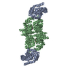
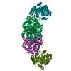
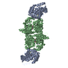
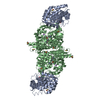
| Title | CRYSTAL STRUCTURES OF MUTANT (BETAK87T) TRYPTOPHAN SYNTHASE ALPHA2 BETA2 COMPLEX WITH LIGANDS BOUND TO THE ACTIVE SITES OF THE ALPHA AND BETA SUBUNITS REVEAL LIGAND-INDUCED CONFORMATIONAL CHANGES | ||||||
 Components Components | (TRYPTOPHAN SYNTHASE) x 2 | ||||||
 Keywords Keywords | LYASE / CARBON-OXYGEN LYASE / TRYPTOPHAN BIOSYNTHESIS / PYRIDOXAL PHOSPHATE | ||||||
| Function / homology |  Function and homology information Function and homology informationtryptophan synthase / tryptophan synthase activity / L-tryptophan biosynthetic process / identical protein binding / cytoplasm / cytosol Similarity search - Function | ||||||
| Biological species |  Salmonella typhimurium (bacteria) Salmonella typhimurium (bacteria) | ||||||
| Method |  X-RAY DIFFRACTION / ISOMORPHOUS WITH PDB ENTRY 1WSY. / Resolution: 1.9 Å X-RAY DIFFRACTION / ISOMORPHOUS WITH PDB ENTRY 1WSY. / Resolution: 1.9 Å | ||||||
 Authors Authors | Rhee, S. / Parris, K.D. / Hyde, C.C. / Ahmed, S.A. / Miles, E.W. / Davies, D.R. | ||||||
 Citation Citation | Journal: Biochemistry / Year: 1997 Title: Crystal structures of a mutant (betaK87T) tryptophan synthase alpha2beta2 complex with ligands bound to the active sites of the alpha- and beta-subunits reveal ligand-induced conformational changes. Authors: Rhee, S. / Parris, K.D. / Hyde, C.C. / Ahmed, S.A. / Miles, E.W. / Davies, D.R. #1: Journal: J.Biol.Chem. / Year: 1993Title: Lysine 87 in the Beta Subunit of Tryptophan Synthase that Forms an Internal Aldimine with Pyridoxal Phosphate Serves Critical Roles in Transimination, Catalysis, and Product Release Authors: Lu, Z. / Nagata, S. / Mcphie, P. / Miles, E.W. | ||||||
| History |
|
- Structure visualization
| Structure viewer | Molecule: MolmilJmol/JSmol |
|---|
- Downloads & links
-Download
| PDBx/mmCIF format | 2tys.cif.gz | 142.8 KB | Display | PDBx/mmCIF format |
|---|---|---|---|---|
| PDB format | pdb2tys.ent.gz | 110.4 KB | Display | PDB format |
| PDBx/mmJSON format | 2tys.json.gz | Tree view | PDBx/mmJSON format | |
| Others |  Other downloads Other downloads |
-Validation report
| Arichive directory | https://data.pdbj.org/pub/pdb/validation_reports/ty/2tysftp://data.pdbj.org/pub/pdb/validation_reports/ty/2tys | HTTPS FTP |
|---|
-Related structure data


-Links
 PDBj
PDBj
- Assembly
| Deposited unit | 
| ||||||||
|---|---|---|---|---|---|---|---|---|---|
| 1 | 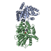
| ||||||||
| Unit cell |
|
-Components
| #1: Protein | Mass: 28698.797 Da / Num. of mol.: 1 / Mutation: CHAIN B, K87T Source method: isolated from a genetically manipulated source Details: L-TRYPTOPHAN BOUND TO THE BETA SUBUNIT IN A FORM OF SCHIFF BASE WITH COENZYME PYRIDOXAL 5'-PHOSPHATE Source: (gene. exp.) Salmonella typhimurium (bacteria) / Cell line: CB149 / Gene: TRPA/TRPB / Plasmid: PSTB7 / Cell line (production host): CB149 / Gene (production host): TRPA/TRPB / Production host: |
|---|---|
| #2: Protein | Mass: 42960.922 Da / Num. of mol.: 1 / Mutation: CHAIN B, K87T Source method: isolated from a genetically manipulated source Details: L-TRYPTOPHAN BOUND TO THE BETA SUBUNIT IN A FORM OF SCHIFF BASE WITH COENZYME PYRIDOXAL 5'-PHOSPHATE Source: (gene. exp.) Salmonella typhimurium (bacteria) / Cell line: CB149 / Gene: TRPA/TRPB / Plasmid: PSTB7 / Cell line (production host): CB149 / Gene (production host): TRPA/TRPB / Production host: |
| #3: Chemical | ChemComp-NA /   Mass: 22.990 Da / Num. of mol.: 1 / Source method: obtained synthetically / Formula: Na Mass: 22.990 Da / Num. of mol.: 1 / Source method: obtained synthetically / Formula: Na |
| #4: Chemical | ChemComp-PLT / [  Mass: 433.352 Da / Num. of mol.: 1 / Source method: obtained synthetically / Formula: C19H20N3O7P Mass: 433.352 Da / Num. of mol.: 1 / Source method: obtained synthetically / Formula: C19H20N3O7P |
| #5: Water | ChemComp-HOH /  Mass: 18.015 Da / Num. of mol.: 358 / Source method: isolated from a natural source / Formula: H2O Mass: 18.015 Da / Num. of mol.: 358 / Source method: isolated from a natural source / Formula: H2O |
-Experimental details
-Experiment
| Experiment | Method: X-RAY DIFFRACTION / Number of used crystals: 2 |
|---|
- Sample preparation
| Crystal | Density Matthews: 2.4 Å3/Da / Density % sol: 48 % | |||||||||||||||||||||||||
|---|---|---|---|---|---|---|---|---|---|---|---|---|---|---|---|---|---|---|---|---|---|---|---|---|---|---|
| Crystal grow | pH: 7.8 Details: 50MM NABICINE (PH 7.8), 1MM NA-EDTA, 0.8-1.5MM SPERMINE, 12% PEG8000 | |||||||||||||||||||||||||
| Crystal grow | *PLUS Method: unknown / Details: pH is adjusted to 7.8 with NaOH | |||||||||||||||||||||||||
| Components of the solutions | *PLUS
|
-Data collection
| Diffraction | Mean temperature: 298 K |
|---|---|
| Diffraction source | Source: ROTATING ANODE / Type: RIGAKU RUH2R / Wavelength: 1.5418 |
| Detector | Type: RIGAKU RAXIS IIC / Detector: IMAGE PLATE / Date: Dec 1, 1992 |
| Radiation | Monochromator: NI FILTER / Monochromatic (M) / Laue (L): M / Scattering type: x-ray |
| Radiation wavelength | Wavelength: 1.5418 Å / Relative weight: 1 |
| Reflection | Highest resolution: 1.9 Å / Num. obs: 50490 / % possible obs: 84.6 % / Observed criterion σ(I): 0 / Redundancy: 2.2 % / Biso Wilson estimate: 22.5 Å2 / Rmerge(I) obs: 0.044 / Net I/σ(I): 18.3 |
| Reflection shell | Resolution: 1.9→2 Å / Mean I/σ(I) obs: 3.7 / % possible all: 46.8 |
| Reflection shell | *PLUS % possible obs: 46.8 % |
- Processing
| Software |
| ||||||||||||||||||||||||||||||||||||||||||||||||||||||||||||||||||||||||||||||||||||
|---|---|---|---|---|---|---|---|---|---|---|---|---|---|---|---|---|---|---|---|---|---|---|---|---|---|---|---|---|---|---|---|---|---|---|---|---|---|---|---|---|---|---|---|---|---|---|---|---|---|---|---|---|---|---|---|---|---|---|---|---|---|---|---|---|---|---|---|---|---|---|---|---|---|---|---|---|---|---|---|---|---|---|---|---|---|
| Refinement | Method to determine structure: ISOMORPHOUS WITH PDB ENTRY 1WSY. Resolution: 1.9→8 Å / σ(F): 2
| ||||||||||||||||||||||||||||||||||||||||||||||||||||||||||||||||||||||||||||||||||||
| Displacement parameters | Biso mean: 29.5 Å2 | ||||||||||||||||||||||||||||||||||||||||||||||||||||||||||||||||||||||||||||||||||||
| Refine analyze | Luzzati coordinate error obs: 0.25 Å / Luzzati d res low obs: 8 Å | ||||||||||||||||||||||||||||||||||||||||||||||||||||||||||||||||||||||||||||||||||||
| Refinement step | Cycle: LAST / Resolution: 1.9→8 Å
| ||||||||||||||||||||||||||||||||||||||||||||||||||||||||||||||||||||||||||||||||||||
| Refine LS restraints |
| ||||||||||||||||||||||||||||||||||||||||||||||||||||||||||||||||||||||||||||||||||||
| Software | *PLUS Name: PROLSQ / Classification: refinement | ||||||||||||||||||||||||||||||||||||||||||||||||||||||||||||||||||||||||||||||||||||
| Refinement | *PLUS % reflection Rfree: 10 % / Rfactor obs: 0.171 / Rfactor Rfree: 0.219 | ||||||||||||||||||||||||||||||||||||||||||||||||||||||||||||||||||||||||||||||||||||
| Solvent computation | *PLUS | ||||||||||||||||||||||||||||||||||||||||||||||||||||||||||||||||||||||||||||||||||||
| Displacement parameters | *PLUS |
