 Movie
Movie Controller

[English] 日本語
 Yorodumi
- PDB-2tgp: THE GEOMETRY OF THE REACTIVE SITE AND OF THE PEPTIDE GROUPS IN TR... -
+ Open data

- Basic information
| Entry | Database: PDB / ID: 2tgp | |||||||||
|---|---|---|---|---|---|---|---|---|---|---|
| Title | THE GEOMETRY OF THE REACTIVE SITE AND OF THE PEPTIDE GROUPS IN TRYPSIN, TRYPSINOGEN AND ITS COMPLEXES WITH INHIBITORS | |||||||||
 Components Components |
| |||||||||
 Keywords Keywords | COMPLEX (PROTEINASE/INHIBITOR) / COMPLEX (PROTEINASE-INHIBITOR) / COMPLEX (PROTEINASE-INHIBITOR) complex | |||||||||
| Function / homology |  Function and homology information Function and homology informationtrypsinogen activation / negative regulation of serine-type endopeptidase activity / sulfate binding / negative regulation of platelet aggregation / potassium channel inhibitor activity / zymogen binding / molecular function inhibitor activity / negative regulation of thrombin-activated receptor signaling pathway / trypsin / serpin family protein binding ...trypsinogen activation / negative regulation of serine-type endopeptidase activity / sulfate binding / negative regulation of platelet aggregation / potassium channel inhibitor activity / zymogen binding / molecular function inhibitor activity / negative regulation of thrombin-activated receptor signaling pathway / trypsin / serpin family protein binding / serine protease inhibitor complex / digestion / serine-type endopeptidase inhibitor activity / protease binding / endopeptidase activity / serine-type endopeptidase activity / calcium ion binding / proteolysis / extracellular space / metal ion binding Similarity search - Function | |||||||||
| Biological species |  | |||||||||
| Method |  X-RAY DIFFRACTION / Resolution: 1.9 Å X-RAY DIFFRACTION / Resolution: 1.9 Å | |||||||||
 Authors Authors | Huber, R. / Bode, W. / Deisenhofer, J. / Schwager, P. | |||||||||
 Citation Citation | Journal: Acta Crystallogr.,Sect.B / Year: 1983 Title: The Geometry of the Reactive Site and of the Peptide Groups in Trypsin, Trypsinogen and its Complexes with Inhibitors Authors: Marquart, M. / Walter, J. / Deisenhofer, J. / Bode, W. / Huber, R. #1: Journal: J.Mol.Biol. / Year: 1979Title: The Transition of Bovine Trypsinogen to a Trypsin-Like State Upon Strong Ligand Binding. II. The Binding of the Pancreatic Trypsin Inhibitor and of Isoleucine-Valine and of Sequentially ...Title: The Transition of Bovine Trypsinogen to a Trypsin-Like State Upon Strong Ligand Binding. II. The Binding of the Pancreatic Trypsin Inhibitor and of Isoleucine-Valine and of Sequentially Related Peptides to Trypsinogen and to P-Guanidinobenzoate-Trypsinogen Authors: Bode, W. #2: Journal: J.Mol.Biol. / Year: 1978Title: The Transition of Bovine Trypsinogen to a Trypsin-Like State Upon Strong Ligand Binding. The Refined Crystal Structures of the Bovine Trypsinogen-Pancreatic Trypsin Inhibitor Complex and of ...Title: The Transition of Bovine Trypsinogen to a Trypsin-Like State Upon Strong Ligand Binding. The Refined Crystal Structures of the Bovine Trypsinogen-Pancreatic Trypsin Inhibitor Complex and of its Ternary Complex with Ile-Val at 1.9 Angstroms Resolution Authors: Bode, W. / Schwager, P. / Huber, R. #3: Journal: Acc.Chem.Res. / Year: 1978Title: Structural Basis of the Activation and Action of Trypsin Authors: Huber, R. / Bode, W. #4: Journal: Biophys.Struct.Mech. / Year: 1975Title: The Structure of the Complex Formed by Bovine Trypsin and Bovine Pancreatic Trypsin Inhibitor. III. Structure of the Anhydro-Trypsin-Inhibitor Complex Authors: Huber, R. / Bode, W. / Kukla, D. / Kohl, U. / Ryan, C.A. #5: Journal: J.Mol.Biol. / Year: 1974Title: Structure of the Complex Formed by Bovine Trypsin and Bovine Pancreatic Trypsin Inhibitor. II. Crystallographic Refinement at 1.9 Angstroms Resolution Authors: Huber, R. / Kukla, D. / Bode, W. / Schwager, P. / Bartels, K. / Deisenhofer, J. / Steigemann, W. | |||||||||
| History |
|
- Structure visualization
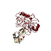
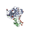
| Structure viewer | Molecule: MolmilJmol/JSmol |
|---|
- Downloads & links
-Download
| PDBx/mmCIF format | 2tgp.cif.gz | 66.3 KB | Display | PDBx/mmCIF format |
|---|---|---|---|---|
| PDB format | pdb2tgp.ent.gz | 49.6 KB | Display | PDB format |
| PDBx/mmJSON format | 2tgp.json.gz | Tree view | PDBx/mmJSON format | |
| Others |  Other downloads Other downloads |
-Validation report
| Summary document | 2tgp_validation.pdf.gz | 433.8 KB | Display | wwPDB validaton report |
|---|---|---|---|---|
| Full document | 2tgp_full_validation.pdf.gz | 440.4 KB | Display | |
| Data in XML | 2tgp_validation.xml.gz | 14.7 KB | Display | |
| Data in CIF | 2tgp_validation.cif.gz | 20.4 KB | Display | |
| Arichive directory | https://data.pdbj.org/pub/pdb/validation_reports/tg/2tgpftp://data.pdbj.org/pub/pdb/validation_reports/tg/2tgp | HTTPS FTP |
-Related structure data
| Similar structure data |
|---|
-Links
 PDBj
PDBj


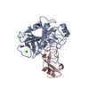
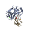
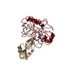
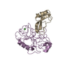
- Assembly
| Deposited unit | 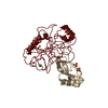
| ||||||||
|---|---|---|---|---|---|---|---|---|---|
| 1 |
| ||||||||
| 2 | 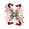
| ||||||||
| 3 | 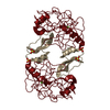
| ||||||||
| 4 | 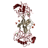
| ||||||||
| Unit cell |
| ||||||||
| Atom site foot note | 1: SEE REMARK 4. | ||||||||
| Components on special symmetry positions |
|
-Components
| #1: Protein | Mass: 24012.953 Da / Num. of mol.: 1 Source method: isolated from a genetically manipulated source Source: (gene. exp.) | ||||||
|---|---|---|---|---|---|---|---|
| #2: Protein | Mass: 6527.568 Da / Num. of mol.: 1 Source method: isolated from a genetically manipulated source Source: (gene. exp.) | ||||||
| #3: Chemical | ChemComp-CA /   Mass: 40.078 Da / Num. of mol.: 1 / Source method: obtained synthetically / Formula: Ca Mass: 40.078 Da / Num. of mol.: 1 / Source method: obtained synthetically / Formula: Ca | ||||||
| #4: Chemical |   Mass: 96.063 Da / Num. of mol.: 2 / Source method: obtained synthetically / Formula: SO4 Mass: 96.063 Da / Num. of mol.: 2 / Source method: obtained synthetically / Formula: SO4#5: Water | ChemComp-HOH / |  Mass: 18.015 Da / Num. of mol.: 138 / Source method: isolated from a natural source / Formula: H2O Mass: 18.015 Da / Num. of mol.: 138 / Source method: isolated from a natural source / Formula: H2OHas protein modification | Y | Sequence details | THE 229 AMINO ACIDS OF TRYPSINOGEN ARE IDENTIFIED BY THE RESIDUE NUMBERS OF THE HOMOLOGOUS ...THE 229 AMINO ACIDS OF TRYPSINOGE | |
-Experimental details
-Experiment
| Experiment | Method: X-RAY DIFFRACTION |
|---|
- Sample preparation
| Crystal | Density Matthews: 3.25 Å3/Da / Density % sol: 62.19 % |
|---|
- Processing
| Refinement | Resolution: 1.9→6.8 Å / Rfactor Rwork: 0.2 Details: THERE IS NO SIGNIFICANT ELECTRON DENSITY IN THE FINAL FOURIER MAP FOR THE N-TERMINUS OF THE ZYMOGEN FROM VAL Z 10 THROUGH GLY Z 18 AND THIS DATA ENTRY CONTAINS NO COORDINATES FOR VAL Z 10 THROUGH LYS Z 15. | ||||||||||||
|---|---|---|---|---|---|---|---|---|---|---|---|---|---|
| Refinement step | Cycle: LAST / Resolution: 1.9→6.8 Å
|
