- PDB-2rr3: Solution structure of the complex between human VAP-A MSP domain ... -
+
Open data
ID or keywords:
Loading...
-
Basic information
Entry
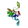
Database: PDB / ID: 2rr3
Title
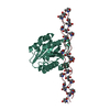
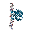
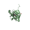
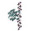
Solution structure of the complex between human VAP-A MSP domain and human OSBP FFAT motif
Components
Oxysterol-binding protein 1
Vesicle-associated membrane protein-associated protein A
Keywords
membrane protein/transport protein / Lipid transport / Transport / PROTEIN-PEPTIDE COMPLEX / major sperm protein domain / protein binding / endoplasmic reticulum / lipid binding / membrane protein-transport protein complex
Function / homology
Function and homology information
phosphatidylinositol-4-phosphate-cholesterol exchange activity / positive regulation of secretory granule organization / FFAT motif binding / : / endoplasmic reticulum-trans-Golgi network membrane contact site / sterol transfer activity / intracellular cholesterol transport / ceramide transport / sphingomyelin biosynthetic process / lipid transfer activity ...phosphatidylinositol-4-phosphate-cholesterol exchange activity / positive regulation of secretory granule organization / FFAT motif binding / : / endoplasmic reticulum-trans-Golgi network membrane contact site / sterol transfer activity / intracellular cholesterol transport / ceramide transport / sphingomyelin biosynthetic process / lipid transfer activity / sterol binding / COPII-coated vesicle budding / sterol transport / host-mediated suppression of viral genome replication / perinuclear endoplasmic reticulum / host-mediated activation of viral genome replication / bile acid biosynthetic process / protein localization to endoplasmic reticulum / Sphingolipid de novo biosynthesis / phospholipid transport / oxysterol binding / cholesterol transport / phosphatidylinositol-4-phosphate binding / azurophil granule membrane / Insertion of tail-anchored proteins into the endoplasmic reticulum membrane / lipid transport / Synthesis of bile acids and bile salts / viral release from host cell / bicellular tight junction / endoplasmic reticulum to Golgi vesicle-mediated transport / protein-membrane adaptor activity / cholesterol homeostasis / positive regulation of insulin secretion involved in cellular response to glucose stimulus / trans-Golgi network / neuron projection development / microtubule cytoskeleton / nuclear membrane / microtubule binding / vesicle / membrane fusion / positive regulation of canonical NF-kappaB signal transduction / cadherin binding / protein heterodimerization activity / Golgi membrane / protein domain specific binding / Neutrophil degranulation / endoplasmic reticulum membrane / perinuclear region of cytoplasm / Golgi apparatus / endoplasmic reticulum / protein homodimerization activity / membrane / plasma membrane / cytoplasm / cytosol Similarity search - Function
Vesicle-associated membrane-protein-associated protein / Major sperm protein (MSP) domain / MSP (Major sperm protein) domain / Major sperm protein (MSP) domain profile. / Oxysterol-binding protein / Oxysterol-binding protein, conserved site / Oxysterol-binding protein superfamily / Oxysterol-binding protein / Oxysterol-binding protein family signature. / PapD-like superfamily ...Vesicle-associated membrane-protein-associated protein / Major sperm protein (MSP) domain / MSP (Major sperm protein) domain / Major sperm protein (MSP) domain profile. / Oxysterol-binding protein / Oxysterol-binding protein, conserved site / Oxysterol-binding protein superfamily / Oxysterol-binding protein / Oxysterol-binding protein family signature. / PapD-like superfamily / PH domain / PH domain profile. / Pleckstrin homology domain. / Pleckstrin homology domain / PH-like domain superfamily / Immunoglobulin-like fold / Immunoglobulins / Immunoglobulin-like / Sandwich / Mainly Beta Similarity search - Domain/homology
Oxysterol-binding protein 1 / Vesicle-associated membrane protein-associated protein A Similarity search - Component
Case, Darden, Cheatham, III, Simmerling, Wang, Duke, Luo, andKollm
refinement
Refinement
Method: molecular dynamics / Software ordinal: 1
NMR representative
Selection criteria: lowest energy
NMR ensemble
Conformer selection criteria: structures with the lowest energy Conformers calculated total number: 300 / Conformers submitted total number: 20 / Representative conformer: 1
+
About Yorodumi
-
News
-
Feb 9, 2022. New format data for meta-information of EMDB entries
New format data for meta-information of EMDB entries
Version 3 of the EMDB header file is now the official format.
The previous official version 1.9 will be removed from the archive.
In the structure databanks used in Yorodumi, some data are registered as the other names, "COVID-19 virus" and "2019-nCoV". Here are the details of the virus and the list of structure data.
Jan 31, 2019. EMDB accession codes are about to change! (news from PDBe EMDB page)
EMDB accession codes are about to change! (news from PDBe EMDB page)
The allocation of 4 digits for EMDB accession codes will soon come to an end. Whilst these codes will remain in use, new EMDB accession codes will include an additional digit and will expand incrementally as the available range of codes is exhausted. The current 4-digit format prefixed with “EMD-” (i.e. EMD-XXXX) will advance to a 5-digit format (i.e. EMD-XXXXX), and so on. It is currently estimated that the 4-digit codes will be depleted around Spring 2019, at which point the 5-digit format will come into force.
The EM Navigator/Yorodumi systems omit the EMD- prefix.
Related info.:Q: What is EMD? / ID/Accession-code notation in Yorodumi/EM Navigator
Yorodumi is a browser for structure data from EMDB, PDB, SASBDB, etc.
This page is also the successor to EM Navigator detail page, and also detail information page/front-end page for Omokage search.
The word "yorodu" (or yorozu) is an old Japanese word meaning "ten thousand". "mi" (miru) is to see.
Related info.:EMDB / PDB / SASBDB / Comparison of 3 databanks / Yorodumi Search / Aug 31, 2016. New EM Navigator & Yorodumi / Yorodumi Papers / Jmol/JSmol / Function and homology information / Changes in new EM Navigator and Yorodumi
 Movie
Movie Controller

 Yorodumi
 Open data
 Basic information
 Components
 Keywords
 Function and homology information
 Homo sapiens (human)
 Authors
 Citation
 Structure visualization
 Downloads & links
 Other downloads


 PDBj
PDBj


 Assembly
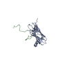

 Sample preparation
 Processing
 NMRPipe