 Movie
Movie Controller

[English] 日本語
 Yorodumi
- PDB-1tzf: X-ray Crystal Structure of alpha-D-glucose-1-phosphate cytidylylt... -
+ Open data

- Basic information
| Entry | Database: PDB / ID: 1tzf | ||||||
|---|---|---|---|---|---|---|---|
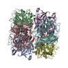
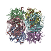
| Title | X-ray Crystal Structure of alpha-D-glucose-1-phosphate cytidylyltransferase from Salmonella typhi | ||||||
 Components Components | Glucose-1-phosphate cytidylyltransferase | ||||||
 Keywords Keywords | TRANSFERASE / nucleotidyltransferase / mixed alpha/beta fold | ||||||
| Function / homology |  Function and homology information Function and homology informationglucose-1-phosphate cytidylyltransferase / glucose-1-phosphate cytidylyltransferase activity / O antigen biosynthetic process / nucleotide binding / metal ion binding Similarity search - Function | ||||||
| Biological species |  Salmonella enterica subsp. enterica serovar Typhi (bacteria) Salmonella enterica subsp. enterica serovar Typhi (bacteria) | ||||||
| Method |  X-RAY DIFFRACTION / SYNCHROTRON / MAD / Resolution: 2.1 Å X-RAY DIFFRACTION / SYNCHROTRON / MAD / Resolution: 2.1 Å | ||||||
 Authors Authors | Koropatkin, N.M. / Holden, H.M. | ||||||
 Citation Citation | Journal: J.Biol.Chem. / Year: 2004 Title: Molecular structure of alpha-D-glucose-1-phosphate cytidylyltransferase from Salmonella typhi Authors: Koropatkin, N.M. / Holden, H.M. | ||||||
| History |
|
- Structure visualization
| Structure viewer | Molecule: MolmilJmol/JSmol |
|---|
- Downloads & links
-Download
| PDBx/mmCIF format | 1tzf.cif.gz | 69.4 KB | Display | PDBx/mmCIF format |
|---|---|---|---|---|
| PDB format | pdb1tzf.ent.gz | 50.5 KB | Display | PDB format |
| PDBx/mmJSON format | 1tzf.json.gz | Tree view | PDBx/mmJSON format | |
| Others |  Other downloads Other downloads |
-Validation report
| Arichive directory | https://data.pdbj.org/pub/pdb/validation_reports/tz/1tzfftp://data.pdbj.org/pub/pdb/validation_reports/tz/1tzf | HTTPS FTP |
|---|
-Related structure data
| Similar structure data |
|---|


-Links
 PDBj
PDBj

- Assembly
| Deposited unit | 
| ||||||||||
|---|---|---|---|---|---|---|---|---|---|---|---|
| 1 | x 6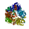
| ||||||||||
| Unit cell |
| ||||||||||
| Components on special symmetry positions |
| ||||||||||
| Details | The biological unit is a hexamer, generated from the monomer in the asymmetric unit by the operations: ROTATION MATRIX: -0.50000 -0.86603 0.00000 0.86603 -0.50000 0.00001 -0.00001 0.00000 1.00000 TRANSLATION VECTOR IN AS 0.00043 0.00017 0.00008 ROTATION MATRIX: -0.50000 0.86602 0.00000 -0.86602 -0.50000 0.00000 0.00000 0.00000 1.00000 TRANSLATION VECTOR IN AS 0.00046 0.00027 0.00009 ROTATION MATRIX: -1.00000 0.00000 0.00000 0.00000 1.00000 0.00001 0.00000 0.00001 -1.00000 TRANSLATION VECTOR IN AS 0.00020 -0.00001 81.14987 ROTATION MATRIX: 0.50000 0.86602 0.00000 0.86602 -0.50000 0.00000 0.00000 0.00000 -1.00000 TRANSLATION VECTOR IN AS 0.00082 0.00015 81.15000 ROTATION MATRIX: 0.50000 -0.86603 0.00000 -0.86603 -0.50000 0.00000 0.00000 0.00000 -1.00000 TRANSLATION VECTOR IN AS 0.00045 0.00058 81.14999 These are the rotation and translation operations to be performed on the original monomer to generate the next five subunits of the complete hexamer 'P 63 2 2' 1 1 0 0 0 1 0 0 0 1 0 0 0 0 0 0 'P 63 2 2' 3 0 -1 0 1 -1 0 0 0 1 0 0 0 0 0 0 'P 63 2 2' 5 -1 1 0 -1 0 0 0 0 1 0 0 0 0 0 0 'P 63 2 2' 7 1 -1 0 0 -1 0 0 0 -1 0 0 0 0 0 0 'P 63 2 2' 9 -1 0 0 -1 1 0 0 0 -1 0 0 0 0 0 0 'P 63 2 2' 11 0 1 0 1 0 0 0 0 -1 0 0 0 0 0 0 'P 63 2 2' 2 1 -1 0 1 0 0 0 0 1 0 0 0 0 1 2 'P 63 2 2' 4 -1 0 0 0 -1 0 0 0 1 0 0 0 0 1 2 'P 63 2 2' 6 0 1 0 -1 1 0 0 0 1 0 0 0 0 1 2 'P 63 2 2' 8 0 -1 0 -1 0 0 0 0 -1 0 0 0 0 1 2 'P 63 2 2' 10 -1 1 0 0 1 0 0 0 -1 0 0 0 0 1 2 'P 63 2 2' 12 1 0 0 1 -1 0 0 0 -1 0 0 0 0 1 2 |
-Components
| #1: Protein | Mass: 29203.598 Da / Num. of mol.: 1 Source method: isolated from a genetically manipulated source Details: complexed with cytidyl-5'-phosphate-glucosyl-6-phosphate Source: (gene. exp.) Salmonella enterica subsp. enterica serovar Typhi (bacteria)Species: Salmonella enterica / Strain: CT18 / Gene: rfbF / Plasmid: pET28a / Production host: References: UniProt: P26396, UniProt: Q8Z5I4*PLUS, glucose-1-phosphate cytidylyltransferase |
|---|---|
| #2: Chemical | ChemComp-MG /   Mass: 24.305 Da / Num. of mol.: 1 / Source method: obtained synthetically / Formula: Mg Mass: 24.305 Da / Num. of mol.: 1 / Source method: obtained synthetically / Formula: Mg |
| #3: Chemical | ChemComp-C5G / [  Mass: 565.317 Da / Num. of mol.: 1 / Source method: obtained synthetically / Formula: C15H25N3O16P2 Mass: 565.317 Da / Num. of mol.: 1 / Source method: obtained synthetically / Formula: C15H25N3O16P2 |
| #4: Water | ChemComp-HOH /  Mass: 18.015 Da / Num. of mol.: 162 / Source method: isolated from a natural source / Formula: H2O Mass: 18.015 Da / Num. of mol.: 162 / Source method: isolated from a natural source / Formula: H2O |
-Experimental details
-Experiment
| Experiment | Method: X-RAY DIFFRACTION / Number of used crystals: 1 |
|---|
- Sample preparation
| Crystal | Density Matthews: 3 Å3/Da / Density % sol: 58.3 % |
|---|---|
| Crystal grow | Temperature: 277 K / Method: vapor diffusion, hanging drop / pH: 5 Details: PEG 4000, Ammonium sulfate, sodium acetate, pH 5.0, VAPOR DIFFUSION, HANGING DROP, temperature 277K |
-Data collection
| Diffraction | Mean temperature: 130 K |
|---|---|
| Diffraction source | Source: SYNCHROTRON / Site: APS  / Beamline: 19-ID / Wavelength: 0.9641 Å / Beamline: 19-ID / Wavelength: 0.9641 Å |
| Detector | Type: SBC-3 / Detector: CCD / Date: Mar 29, 2004 |
| Radiation | Monochromator: Double crystal Si-111 / Protocol: MAD / Monochromatic (M) / Laue (L): M / Scattering type: x-ray |
| Radiation wavelength | Wavelength: 0.9641 Å / Relative weight: 1 |
| Reflection | Resolution: 2.1→50 Å / Num. all: 22401 / Num. obs: 22358 / % possible obs: 98.5 % / Observed criterion σ(F): 0 / Observed criterion σ(I): 0 / Redundancy: 14.1 % / Rsym value: 0.067 / Net I/σ(I): 65.3 |
| Reflection shell | Resolution: 2.1→2.18 Å / Redundancy: 5.4 % / Mean I/σ(I) obs: 9.9 / Num. unique all: 2163 / Rsym value: 0.176 / % possible all: 98.1 |
- Processing
| Software |
| |||||||||||||||||||||||||
|---|---|---|---|---|---|---|---|---|---|---|---|---|---|---|---|---|---|---|---|---|---|---|---|---|---|---|
| Refinement | Method to determine structure: MAD / Resolution: 2.1→50 Å / Cross valid method: THROUGHOUT / σ(F): 0
| |||||||||||||||||||||||||
| Refinement step | Cycle: LAST / Resolution: 2.1→50 Å
| |||||||||||||||||||||||||
| Refine LS restraints |
|
