 Movie
Movie Controller

[English] 日本語
 Yorodumi
- PDB-5ayd: Crystal structure of Ruminococcus albus beta-(1,4)-mannooligosacc... -
+ Open data

- Basic information
| Entry | Database: PDB / ID: 5ayd | ||||||
|---|---|---|---|---|---|---|---|
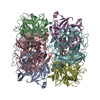
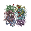
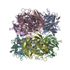
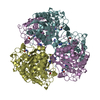
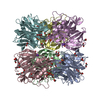
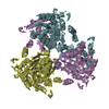
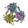
| Title | Crystal structure of Ruminococcus albus beta-(1,4)-mannooligosaccharide phosphorylase (RaMP2) in complexes with phosphate | ||||||
 Components Components | Beta-1,4-mannooligosaccharide phosphorylase | ||||||
 Keywords Keywords | TRANSFERASE / Glycoside hydrolase family 130 | ||||||
| Function / homology |  Function and homology information Function and homology informationbeta-1,4-mannooligosaccharide phosphorylase / glycosyltransferase activity / cell wall organization Similarity search - Function | ||||||
| Biological species |  Ruminococcus albus (bacteria) Ruminococcus albus (bacteria) | ||||||
| Method |  X-RAY DIFFRACTION / SYNCHROTRON / MOLECULAR REPLACEMENT / Resolution: 2.3 Å X-RAY DIFFRACTION / SYNCHROTRON / MOLECULAR REPLACEMENT / Resolution: 2.3 Å | ||||||
 Authors Authors | Ye, Y. / Saburi, W. / Kato, K. / Yao, M. | ||||||
 Citation Citation | Journal: Febs Lett. / Year: 2016 Title: Structural insights into the difference in substrate recognition of two mannoside phosphorylases from two GH130 subfamilies. Authors: Ye, Y. / Saburi, W. / Odaka, R. / Kato, K. / Sakurai, N. / Komoda, K. / Nishimoto, M. / Kitaoka, M. / Mori, H. / Yao, M. | ||||||
| History |
|
- Structure visualization
| Structure viewer | Molecule: MolmilJmol/JSmol |
|---|
- Downloads & links
-Download
| PDBx/mmCIF format | 5ayd.cif.gz | 424.1 KB | Display | PDBx/mmCIF format |
|---|---|---|---|---|
| PDB format | pdb5ayd.ent.gz | 346.5 KB | Display | PDB format |
| PDBx/mmJSON format | 5ayd.json.gz | Tree view | PDBx/mmJSON format | |
| Others |  Other downloads Other downloads |
-Validation report
| Arichive directory | https://data.pdbj.org/pub/pdb/validation_reports/ay/5aydftp://data.pdbj.org/pub/pdb/validation_reports/ay/5ayd | HTTPS FTP |
|---|
-Related structure data


-Links
 PDBj
PDBj- Assembly
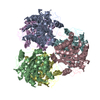
| Deposited unit | 
| ||||||||
|---|---|---|---|---|---|---|---|---|---|
| 1 |
| ||||||||
| Unit cell |
|
-Components
| #1: Protein | Mass: 38352.293 Da / Num. of mol.: 6 / Fragment: UNP residues 1-335 Source method: isolated from a genetically manipulated source Source: (gene. exp.) Ruminococcus albus (strain ATCC 27210 / DSM 20455 / JCM 14654 / NCDO 2250 / 7) (bacteria)Strain: ATCC 27210 / DSM 20455 / JCM 14654 / NCDO 2250 / 7 / Gene: Rumal_0099 / Production host: References: UniProt: E6UBR9, beta-1,4-mannooligosaccharide phosphorylase #2: Chemical | ChemComp-PO4 /   Mass: 94.971 Da / Num. of mol.: 6 / Source method: obtained synthetically / Formula: PO4 Mass: 94.971 Da / Num. of mol.: 6 / Source method: obtained synthetically / Formula: PO4#3: Water | ChemComp-HOH / |  Mass: 18.015 Da / Num. of mol.: 1212 / Source method: isolated from a natural source / Formula: H2O Mass: 18.015 Da / Num. of mol.: 1212 / Source method: isolated from a natural source / Formula: H2O |
|---|
-Experimental details
-Experiment
| Experiment | Method: X-RAY DIFFRACTION |
|---|
- Sample preparation
| Crystal | Density Matthews: 2.72 Å3/Da / Density % sol: 54.84 % |
|---|---|
| Crystal grow | Temperature: 293 K / Method: vapor diffusion, sitting drop Details: 0.2M Ammonium phosphate, 0.1M Tris-HCl buffer, 50%(v/v) MPD |
-Data collection
| Diffraction | Mean temperature: 100 K |
|---|---|
| Diffraction source | Source: SYNCHROTRON / Site: Photon Factory  / Beamline: AR-NW12A / Wavelength: 1 Å / Beamline: AR-NW12A / Wavelength: 1 Å |
| Detector | Type: ADSC QUANTUM 210r / Detector: CCD / Date: Nov 4, 2013 |
| Radiation | Protocol: SINGLE WAVELENGTH / Monochromatic (M) / Laue (L): M / Scattering type: x-ray |
| Radiation wavelength | Wavelength: 1 Å / Relative weight: 1 |
| Reflection | Resolution: 2.3→44.76 Å / Num. obs: 109233 / % possible obs: 99.4 % / Redundancy: 3.8 % / Net I/σ(I): 9.75 |
- Processing
| Software |
| |||||||||||||||||||||||||||||||||||||||||||||||||||||||||||||||||||||||||||||||||||||||||||||||||||||||||||||||||||||||||||||||||||||||||||||||||||||||||||||||||||||||||||||||||||||||||||||||||||||||||||||||||||||||||
|---|---|---|---|---|---|---|---|---|---|---|---|---|---|---|---|---|---|---|---|---|---|---|---|---|---|---|---|---|---|---|---|---|---|---|---|---|---|---|---|---|---|---|---|---|---|---|---|---|---|---|---|---|---|---|---|---|---|---|---|---|---|---|---|---|---|---|---|---|---|---|---|---|---|---|---|---|---|---|---|---|---|---|---|---|---|---|---|---|---|---|---|---|---|---|---|---|---|---|---|---|---|---|---|---|---|---|---|---|---|---|---|---|---|---|---|---|---|---|---|---|---|---|---|---|---|---|---|---|---|---|---|---|---|---|---|---|---|---|---|---|---|---|---|---|---|---|---|---|---|---|---|---|---|---|---|---|---|---|---|---|---|---|---|---|---|---|---|---|---|---|---|---|---|---|---|---|---|---|---|---|---|---|---|---|---|---|---|---|---|---|---|---|---|---|---|---|---|---|---|---|---|---|---|---|---|---|---|---|---|---|---|---|---|---|---|---|---|---|
| Refinement | Method to determine structure: MOLECULAR REPLACEMENT / Resolution: 2.3→44.757 Å / SU ML: 0.27 / Cross valid method: FREE R-VALUE / σ(F): 1.37 / Phase error: 20.78 / Stereochemistry target values: ML
| |||||||||||||||||||||||||||||||||||||||||||||||||||||||||||||||||||||||||||||||||||||||||||||||||||||||||||||||||||||||||||||||||||||||||||||||||||||||||||||||||||||||||||||||||||||||||||||||||||||||||||||||||||||||||
| Solvent computation | Shrinkage radii: 0.9 Å / VDW probe radii: 1.11 Å / Solvent model: FLAT BULK SOLVENT MODEL | |||||||||||||||||||||||||||||||||||||||||||||||||||||||||||||||||||||||||||||||||||||||||||||||||||||||||||||||||||||||||||||||||||||||||||||||||||||||||||||||||||||||||||||||||||||||||||||||||||||||||||||||||||||||||
| Refinement step | Cycle: LAST / Resolution: 2.3→44.757 Å
| |||||||||||||||||||||||||||||||||||||||||||||||||||||||||||||||||||||||||||||||||||||||||||||||||||||||||||||||||||||||||||||||||||||||||||||||||||||||||||||||||||||||||||||||||||||||||||||||||||||||||||||||||||||||||
| Refine LS restraints |
| |||||||||||||||||||||||||||||||||||||||||||||||||||||||||||||||||||||||||||||||||||||||||||||||||||||||||||||||||||||||||||||||||||||||||||||||||||||||||||||||||||||||||||||||||||||||||||||||||||||||||||||||||||||||||
| LS refinement shell |
|
