 Movie
Movie Controller

[English] 日本語
 Yorodumi
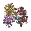
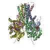
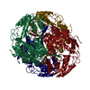
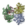
- PDB-1wvc: alpha-D-glucose-1-phosphate cytidylyltransferase complexed with CTP -
+ Open data

- Basic information
| Entry | Database: PDB / ID: 1wvc | ||||||
|---|---|---|---|---|---|---|---|
| Title | alpha-D-glucose-1-phosphate cytidylyltransferase complexed with CTP | ||||||
 Components Components | Glucose-1-phosphate cytidylyltransferase | ||||||
 Keywords Keywords | TRANSFERASE / CDP-glucose pyrophosphorylase / nucleotidyltransferase | ||||||
| Function / homology |  Function and homology information Function and homology informationglucose-1-phosphate cytidylyltransferase / glucose-1-phosphate cytidylyltransferase activity / O antigen biosynthetic process / nucleotide binding / metal ion binding Similarity search - Function | ||||||
| Biological species |  Salmonella enterica subsp. enterica serovar Typhi (bacteria) Salmonella enterica subsp. enterica serovar Typhi (bacteria) | ||||||
| Method |  X-RAY DIFFRACTION / SYNCHROTRON / MOLECULAR REPLACEMENT / Resolution: 2.5 Å X-RAY DIFFRACTION / SYNCHROTRON / MOLECULAR REPLACEMENT / Resolution: 2.5 Å | ||||||
 Authors Authors | Koropatkin, N.M. / Cleland, W.W. / Holden, H.M. | ||||||
 Citation Citation | Journal: J.Biol.Chem. / Year: 2005 Title: Kinetic and structural analysis of alpha-D-Glucose-1-phosphate cytidylyltransferase from Salmonella typhi. Authors: Koropatkin, N.M. / Cleland, W.W. / Holden, H.M. | ||||||
| History |
|
- Structure visualization
| Structure viewer | Molecule: MolmilJmol/JSmol |
|---|
- Downloads & links
-Download
| PDBx/mmCIF format | 1wvc.cif.gz | 68.2 KB | Display | PDBx/mmCIF format |
|---|---|---|---|---|
| PDB format | pdb1wvc.ent.gz | 49 KB | Display | PDB format |
| PDBx/mmJSON format | 1wvc.json.gz | Tree view | PDBx/mmJSON format | |
| Others |  Other downloads Other downloads |
-Validation report
| Arichive directory | https://data.pdbj.org/pub/pdb/validation_reports/wv/1wvcftp://data.pdbj.org/pub/pdb/validation_reports/wv/1wvc | HTTPS FTP |
|---|
-Related structure data
| Related structure data |  1tzfS S: Starting model for refinement |
|---|---|
| Similar structure data |

-Links
 PDBj
PDBj


- Assembly
| Deposited unit | 
| ||||||||
|---|---|---|---|---|---|---|---|---|---|
| 1 | x 6
| ||||||||
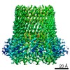
| Unit cell |
|
-Components
| #1: Protein | Mass: 29203.598 Da / Num. of mol.: 1 Source method: isolated from a genetically manipulated source Source: (gene. exp.) Salmonella enterica subsp. enterica serovar Typhi (bacteria)Species: Salmonella enterica / Strain: CT18 / Gene: rfbF / Plasmid: pET-28a / Production host: References: UniProt: P26396, UniProt: Q8Z5I4*PLUS, glucose-1-phosphate cytidylyltransferase | ||||||
|---|---|---|---|---|---|---|---|
| #2: Chemical |   Mass: 24.305 Da / Num. of mol.: 2 / Source method: obtained synthetically / Formula: Mg Mass: 24.305 Da / Num. of mol.: 2 / Source method: obtained synthetically / Formula: Mg#3: Chemical | ChemComp-NI / |   Mass: 58.693 Da / Num. of mol.: 1 / Source method: obtained synthetically / Formula: Ni Mass: 58.693 Da / Num. of mol.: 1 / Source method: obtained synthetically / Formula: Ni#4: Chemical | ChemComp-CTP / |   Mass: 483.156 Da / Num. of mol.: 1 / Source method: obtained synthetically / Formula: C9H16N3O14P3 Mass: 483.156 Da / Num. of mol.: 1 / Source method: obtained synthetically / Formula: C9H16N3O14P3#5: Water | ChemComp-HOH / |  Mass: 18.015 Da / Num. of mol.: 91 / Source method: isolated from a natural source / Formula: H2O Mass: 18.015 Da / Num. of mol.: 91 / Source method: isolated from a natural source / Formula: H2O |
-Experimental details
-Experiment
| Experiment | Method: X-RAY DIFFRACTION / Number of used crystals: 1 |
|---|
- Sample preparation
| Crystal | Density Matthews: 3 Å3/Da / Density % sol: 58.3 % |
|---|---|
| Crystal grow | Temperature: 298 K / Method: vapor diffusion, hanging drop / pH: 7 Details: PEG 400, magnesium chloride, HEPES, pH 7.0, VAPOR DIFFUSION, HANGING DROP, temperature 298.0K |
-Data collection
| Diffraction | Mean temperature: 130 K |
|---|---|
| Diffraction source | Source: SYNCHROTRON / Site: APS  / Beamline: 32-ID / Wavelength: 0.9641 Å / Beamline: 32-ID / Wavelength: 0.9641 Å |
| Detector | Type: MARRESEARCH / Detector: CCD / Date: Aug 6, 2004 |
| Radiation | Monochromator: Diamond 111 / Protocol: SINGLE WAVELENGTH / Monochromatic (M) / Laue (L): M / Scattering type: x-ray |
| Radiation wavelength | Wavelength: 0.9641 Å / Relative weight: 1 |
| Reflection | Resolution: 2.5→25 Å / Num. all: 11780 / Num. obs: 11780 / % possible obs: 97.4 % / Observed criterion σ(F): 0 / Observed criterion σ(I): 0 / Redundancy: 9.5 % / Rsym value: 0.062 / Net I/σ(I): 26.4 |
| Reflection shell | Resolution: 2.5→2.59 Å / Redundancy: 5.3 % / Mean I/σ(I) obs: 3.1 / Num. unique all: 1064 / Rsym value: 0.274 / % possible all: 90.3 |
- Processing
| Software |
| |||||||||||||||||||||||||
|---|---|---|---|---|---|---|---|---|---|---|---|---|---|---|---|---|---|---|---|---|---|---|---|---|---|---|
| Refinement | Method to determine structure: MOLECULAR REPLACEMENT Starting model: PDB entry 1TZF Resolution: 2.5→25 Å / σ(F): 0.5
| |||||||||||||||||||||||||
| Refinement step | Cycle: LAST / Resolution: 2.5→25 Å
| |||||||||||||||||||||||||
| Refine LS restraints |
|
