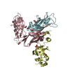
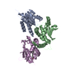
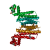
BIOMOLECULE: 1 SEE REMARK 350 FOR THE AUTHOR PROVIDED AND PROGRAM GENERATED ASSEMBLY INFORMATION ... BIOMOLECULE: 1 SEE REMARK 350 FOR THE AUTHOR PROVIDED AND PROGRAM GENERATED ASSEMBLY INFORMATION FOR THE STRUCTURE IN THIS ENTRY. THE REMARK MAY ALSO PROVIDE INFORMATION ON BURIED SURFACE AREA. SIZE EXCLUSION CHROMATOGRAPHY SUPPORTS THE ASSIGNMENT OF A DIMER AS THE SIGNIFICANT OLIGOMERIZATION STATE.
Remark 999
SEQUENCE THE CONSTRUCT WAS EXPRESSED WITH A PURIFICATION TAG MGSDKIHHHHHHENLYFQG. THE TAG WAS ... SEQUENCE THE CONSTRUCT WAS EXPRESSED WITH A PURIFICATION TAG MGSDKIHHHHHHENLYFQG. THE TAG WAS REMOVED WITH TEV PROTEASE LEAVING ONLY A GLYCINE (0) FOLLOWED BY THE TARGET SEQUENCE.
Monochromator: Single crystal Si(111) bent (horizontal focusing) Protocol: MAD / Monochromatic (M) / Laue (L): M / Scattering type: x-ray
Radiation wavelength
ID
Wavelength (Å)
Relative weight
1
0.91837
1
2
0.97925
1
3
0.97901
1
Reflection
Resolution: 1.8→29.709 Å / Num. obs: 47914 / % possible obs: 96.7 % / Observed criterion σ(I): -3 / Biso Wilson estimate: 27.39 Å2 / Rmerge(I) obs: 0.034 / Net I/σ(I): 14.01
Reflection shell
Resolution (Å)
Rmerge(I) obs
Mean I/σ(I) obs
Num. measured obs
Num. unique obs
Diffraction-ID
% possible all
1.8-1.86
0.432
2
12756
8152
1
93.9
1.86-1.94
0.328
2.7
15344
9736
1
98
1.94-2.03
0.22
3.8
14572
9220
1
98.3
2.03-2.13
0.149
5.6
13581
8548
1
98.2
2.13-2.27
0.099
8.1
15156
9539
1
98.3
2.27-2.44
0.07
11.1
14152
8782
1
98.3
2.44-2.69
0.05
14.4
14880
9230
1
97.8
2.69-3.07
0.033
20.9
14470
8814
1
97.1
3.07-3.87
0.019
31.9
14916
8919
1
95.9
3.87-29.709
0.016
41.2
14751
8519
1
91.3
-
Phasing
Phasing
Method: MAD
-
Processing
Software
Name
Version
Classification
NB
REFMAC
5.2.0019
refinement
PHENIX
refinement
SHELX
phasing
MolProbity
3beta29
modelbuilding
XSCALE
datascaling
PDB_EXTRACT
3
dataextraction
MAR345
CCD
datacollection
XDS
datareduction
SHELXD
phasing
autoSHARP
phasing
Refinement
Method to determine structure: MAD / Resolution: 1.8→29.709 Å / Cor.coef. Fo:Fc: 0.966 / Cor.coef. Fo:Fc free: 0.95 / SU B: 5.258 / SU ML: 0.081 / TLS residual ADP flag: LIKELY RESIDUAL / Cross valid method: THROUGHOUT / σ(F): 0 / ESU R: 0.111 / ESU R Free: 0.107 Stereochemistry target values: MAXIMUM LIKELIHOOD WITH PHASES Details: 1. HYDROGENS HAVE BEEN ADDED IN THE RIDING POSITIONS. 2. ATOM RECORDS CONTAIN RESIDUAL B FACTORS ONLY. 3. A MET-INHIBITION PROTOCOL WAS USED FOR SELENOMETHIONINE INCORPORATION DURING PROTEIN ...Details: 1. HYDROGENS HAVE BEEN ADDED IN THE RIDING POSITIONS. 2. ATOM RECORDS CONTAIN RESIDUAL B FACTORS ONLY. 3. A MET-INHIBITION PROTOCOL WAS USED FOR SELENOMETHIONINE INCORPORATION DURING PROTEIN EXPRESSION. THE OCCUPANCY OF THE SE ATOMS IN THE MSE RESIDUES WAS REDUCED TO 0.75 FOR THE REDUCED SCATTERING POWER DUE TO PARTIAL S-MET INCORPORATION. 4. RESIDUES 1-4 IN BOTH CHAINS, 205-211 IN CHAIN A, AND 209-211 IN CHAIN B ARE DISORDERED AND NOT INCLUDED IN THE MODEL. 5. EDO MOLECULES FROM THE CRYO SOLUTION ARE MODELED. 6. THERE ARE UNEXPLAINED DENSITIES NEAR GLU-121 SUGGESTING A MG ION COORDINATED BY WATERS.
Rfactor
Num. reflection
% reflection
Selection details
Rfree
0.206
2413
5 %
RANDOM
Rwork
0.177
-
-
-
obs
0.178
47877
98.77 %
-
Solvent computation
Ion probe radii: 0.8 Å / Shrinkage radii: 0.8 Å / VDW probe radii: 1.2 Å / Solvent model: BABINET MODEL WITH MASK
Displacement parameters
Biso mean: 34.359 Å2
Baniso -1
Baniso -2
Baniso -3
1-
2.13 Å2
0 Å2
0 Å2
2-
-
-0.23 Å2
0 Å2
3-
-
-
-1.89 Å2
Refinement step
Cycle: LAST / Resolution: 1.8→29.709 Å
Protein
Nucleic acid
Ligand
Solvent
Total
Num. atoms
3133
0
58
353
3544
Refine LS restraints
Refine-ID
Type
Dev ideal
Dev ideal target
Number
X-RAY DIFFRACTION
r_bond_refined_d
0.015
0.022
3342
X-RAY DIFFRACTION
r_bond_other_d
0.004
0.02
2311
X-RAY DIFFRACTION
r_angle_refined_deg
1.422
1.964
4517
X-RAY DIFFRACTION
r_angle_other_deg
1.427
3
5571
X-RAY DIFFRACTION
r_dihedral_angle_1_deg
3.534
5
438
X-RAY DIFFRACTION
r_dihedral_angle_2_deg
26.8
23.072
166
X-RAY DIFFRACTION
r_dihedral_angle_3_deg
11.949
15
558
X-RAY DIFFRACTION
r_dihedral_angle_4_deg
15.251
15
36
X-RAY DIFFRACTION
r_chiral_restr
0.094
0.2
498
X-RAY DIFFRACTION
r_gen_planes_refined
0.006
0.02
3796
X-RAY DIFFRACTION
r_gen_planes_other
0.003
0.02
736
X-RAY DIFFRACTION
r_nbd_refined
0.192
0.2
716
X-RAY DIFFRACTION
r_nbd_other
0.145
0.2
2378
X-RAY DIFFRACTION
r_nbtor_refined
0.162
0.2
1645
X-RAY DIFFRACTION
r_nbtor_other
0.075
0.2
1640
X-RAY DIFFRACTION
r_xyhbond_nbd_refined
0.124
0.2
280
X-RAY DIFFRACTION
r_symmetry_vdw_refined
0.098
0.2
12
X-RAY DIFFRACTION
r_symmetry_vdw_other
0.198
0.2
66
X-RAY DIFFRACTION
r_symmetry_hbond_refined
0.121
0.2
25
X-RAY DIFFRACTION
r_mcbond_it
1.777
3
2131
X-RAY DIFFRACTION
r_mcbond_other
0.411
3
839
X-RAY DIFFRACTION
r_mcangle_it
2.571
5
3290
X-RAY DIFFRACTION
r_scbond_it
4.459
8
1379
X-RAY DIFFRACTION
r_scangle_it
6.269
11
1209
LS refinement shell
Resolution: 1.8→1.846 Å / Total num. of bins used: 20
Rfactor
Num. reflection
% reflection
Rfree
0.321
161
-
Rwork
0.25
3271
-
all
-
3432
-
obs
-
-
96.51 %
Refinement TLS params.
Method: refined / Refine-ID: X-RAY DIFFRACTION
ID
L11 (°2)
L12 (°2)
L13 (°2)
L22 (°2)
L23 (°2)
L33 (°2)
S11 (Å °)
S12 (Å °)
S13 (Å °)
S21 (Å °)
S22 (Å °)
S23 (Å °)
S31 (Å °)
S32 (Å °)
S33 (Å °)
T11 (Å2)
T12 (Å2)
T13 (Å2)
T22 (Å2)
T23 (Å2)
T33 (Å2)
Origin x (Å)
Origin y (Å)
Origin z (Å)
1
1.2931
-0.4127
-1.0422
0.9875
0.3523
1.838
-0.0269
0.0519
0.0904
-0.0767
0.1236
-0.1979
0.0703
0.0717
-0.0967
-0.2051
-0.0177
-0.011
-0.1563
-0.0313
-0.1096
14.144
18.224
20.71
2
0.8706
-0.4458
0.4644
1.3377
0.1727
2.4724
-0.0276
0.0119
0.0059
-0.125
0.0618
0.1824
-0.1444
-0.2241
-0.0342
-0.1968
-0.0124
0.0008
-0.176
0.0074
-0.1369
-13.368
29.612
22.246
Refinement TLS group
ID
Refine-ID
Refine TLS-ID
Auth asym-ID
Label asym-ID
Auth seq-ID
Label seq-ID
1
X-RAY DIFFRACTION
1
A
A
5 - 204
6 - 205
2
X-RAY DIFFRACTION
2
B
B
5 - 208
6 - 209
+
About Yorodumi
-
News
-
Feb 9, 2022. New format data for meta-information of EMDB entries
New format data for meta-information of EMDB entries
Version 3 of the EMDB header file is now the official format.
The previous official version 1.9 will be removed from the archive.
In the structure databanks used in Yorodumi, some data are registered as the other names, "COVID-19 virus" and "2019-nCoV". Here are the details of the virus and the list of structure data.
Jan 31, 2019. EMDB accession codes are about to change! (news from PDBe EMDB page)
EMDB accession codes are about to change! (news from PDBe EMDB page)
The allocation of 4 digits for EMDB accession codes will soon come to an end. Whilst these codes will remain in use, new EMDB accession codes will include an additional digit and will expand incrementally as the available range of codes is exhausted. The current 4-digit format prefixed with “EMD-” (i.e. EMD-XXXX) will advance to a 5-digit format (i.e. EMD-XXXXX), and so on. It is currently estimated that the 4-digit codes will be depleted around Spring 2019, at which point the 5-digit format will come into force.
The EM Navigator/Yorodumi systems omit the EMD- prefix.
Related info.:Q: What is EMD? / ID/Accession-code notation in Yorodumi/EM Navigator
Yorodumi is a browser for structure data from EMDB, PDB, SASBDB, etc.
This page is also the successor to EM Navigator detail page, and also detail information page/front-end page for Omokage search.
The word "yorodu" (or yorozu) is an old Japanese word meaning "ten thousand". "mi" (miru) is to see.
Related info.:EMDB / PDB / SASBDB / Comparison of 3 databanks / Yorodumi Search / Aug 31, 2016. New EM Navigator & Yorodumi / Yorodumi Papers / Jmol/JSmol / Function and homology information / Changes in new EM Navigator and Yorodumi
 Movie
Movie Controller

 Yorodumi
 Open data
 Basic information
 Components
 Keywords
 Function and homology information
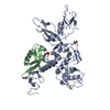
 Novosphingobium aromaticivorans (bacteria)
 X-RAY DIFFRACTION /
  Authors
 Citation
 Structure visualization
 Downloads & links
 Other downloads
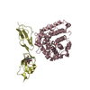


 PDBj
PDBj

 Assembly

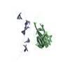
 Mass: 35.453 Da / Num. of mol.: 2 / Source method: obtained synthetically / Formula: Cl
 Mass: 62.068 Da / Num. of mol.: 14 / Source method: obtained synthetically / Formula: C2H6O2
 Mass: 18.015 Da / Num. of mol.: 353 / Source method: isolated from a natural source / Formula: H2O
 Sample preparation
 / Beamline: BL11-1 / Wavelength: 0.91837, 0.97925, 0.97901
 Processing