 Movie
Movie Controller


+ Open data

- Basic information
| Entry | Database: PDB / ID: 1d4v | ||||||
|---|---|---|---|---|---|---|---|
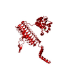
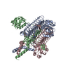
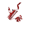
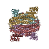
| Title | Crystal structure of trail-DR5 complex | ||||||
 Components Components |
| ||||||
 Keywords Keywords | APOPTOSIS / LIGAND-RECEPTOR COMPLEX / TRIMERIC JELLY-ROLL / TNF-R SUPERFAMILY | ||||||
| Function / homology |  Function and homology information Function and homology informationTRAIL receptor activity / TRAIL binding / TRAIL signaling / TRAIL-activated apoptotic signaling pathway / Regulation by c-FLIP / CASP8 activity is inhibited / Dimerization of procaspase-8 / Caspase activation via Death Receptors in the presence of ligand / activation of NF-kappaB-inducing kinase activity / defense response to tumor cell ...TRAIL receptor activity / TRAIL binding / TRAIL signaling / TRAIL-activated apoptotic signaling pathway / Regulation by c-FLIP / CASP8 activity is inhibited / Dimerization of procaspase-8 / Caspase activation via Death Receptors in the presence of ligand / activation of NF-kappaB-inducing kinase activity / defense response to tumor cell / tumor necrosis factor receptor binding / TP53 Regulates Transcription of Death Receptors and Ligands / positive regulation of extrinsic apoptotic signaling pathway / RIPK1-mediated regulated necrosis / positive regulation of release of cytochrome c from mitochondria / intrinsic apoptotic signaling pathway in response to endoplasmic reticulum stress / extrinsic apoptotic signaling pathway via death domain receptors / response to endoplasmic reticulum stress / cytokine activity / Cell surface interactions at the vascular wall / cellular response to mechanical stimulus / cell-cell signaling / signaling receptor activity / regulation of apoptotic process / positive regulation of canonical NF-kappaB signal transduction / cell surface receptor signaling pathway / immune response / positive regulation of apoptotic process / signaling receptor binding / apoptotic process / cell surface / signal transduction / : / extracellular exosome / extracellular region / zinc ion binding / identical protein binding / plasma membrane Similarity search - Function | ||||||
| Biological species |  Homo sapiens (human) Homo sapiens (human) | ||||||
| Method |  X-RAY DIFFRACTION / SYNCHROTRON / Resolution: 2.2 Å X-RAY DIFFRACTION / SYNCHROTRON / Resolution: 2.2 Å | ||||||
 Authors Authors | Mongkolsapaya, J. / Grimes, J.M. / Stuart, D.I. / Jones, E.Y. / Screaton, G.R. | ||||||
 Citation Citation | Journal: Nat.Struct.Biol. / Year: 1999 Title: Structure of the TRAIL-DR5 complex reveals mechanisms conferring specificity in apoptotic initiation Authors: Mongkolsapaya, J. / Grimes, J.M. / Chen, N. / Xu, X.N. / Stuart, D.I. / Jones, E.Y. / Screaton, G.R. | ||||||
| History |
|
- Structure visualization
| Structure viewer | Molecule: MolmilJmol/JSmol |
|---|
- Downloads & links
-Download
| PDBx/mmCIF format | 1d4v.cif.gz | 71.9 KB | Display | PDBx/mmCIF format |
|---|---|---|---|---|
| PDB format | pdb1d4v.ent.gz | 54 KB | Display | PDB format |
| PDBx/mmJSON format | 1d4v.json.gz | Tree view | PDBx/mmJSON format | |
| Others |  Other downloads Other downloads |
-Validation report
| Arichive directory | https://data.pdbj.org/pub/pdb/validation_reports/d4/1d4vftp://data.pdbj.org/pub/pdb/validation_reports/d4/1d4v | HTTPS FTP |
|---|
-Related structure data
| Similar structure data |
|---|


-Links
 PDBj
PDBj


- Assembly
| Deposited unit | 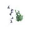
| ||||||||
|---|---|---|---|---|---|---|---|---|---|
| 1 | 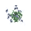
| ||||||||
| Unit cell |
|
-Components
| #1: Protein | Mass: 18921.166 Da / Num. of mol.: 1 / Fragment: SINGLE SUBUNIT Source method: isolated from a genetically manipulated source Source: (gene. exp.) Homo sapiens (human) / Plasmid: PET-9C / Production host:  |
|---|---|
| #2: Protein | Mass: 13143.660 Da / Num. of mol.: 1 / Fragment: EXTRACELLULAR REGION Source method: isolated from a genetically manipulated source Source: (gene. exp.) Homo sapiens (human) / Gene: TRANSIENT EXPRESSION AS IG FUSION PROTEIN / Cell (production host): 293T CELLS / Production host: Homo sapiens (human) / References: GenBank: 2338420, UniProt: O14763*PLUS |
| #3: Water | ChemComp-HOH /  Mass: 18.015 Da / Num. of mol.: 154 / Source method: isolated from a natural source / Formula: H2O Mass: 18.015 Da / Num. of mol.: 154 / Source method: isolated from a natural source / Formula: H2O |
| Has protein modification | Y |
-Experimental details
-Experiment
| Experiment | Method: X-RAY DIFFRACTION / Number of used crystals: 1 |
|---|
- Sample preparation
| Crystal | Density Matthews: 2.87 Å3/Da / Density % sol: 57.1 % | ||||||||||||||||||||||||||||||||||||
|---|---|---|---|---|---|---|---|---|---|---|---|---|---|---|---|---|---|---|---|---|---|---|---|---|---|---|---|---|---|---|---|---|---|---|---|---|---|
| Crystal grow | Temperature: 293 K / Method: vapor diffusion, sitting drop / pH: 8 Details: 25% ETHYLENE GLYCOL, 0.1% N-OCTYL-BETA-GLUCOSIDE, pH 8, VAPOR DIFFUSION, SITTING DROP, temperature 20K | ||||||||||||||||||||||||||||||||||||
| Crystal grow | *PLUS | ||||||||||||||||||||||||||||||||||||
| Components of the solutions | *PLUS
|
-Data collection
| Diffraction | Mean temperature: 100 K |
|---|---|
| Diffraction source | Source: SYNCHROTRON / Site: SRS  / Beamline: PX9.6 / Wavelength: 0.87 / Beamline: PX9.6 / Wavelength: 0.87 |
| Detector | Type: ADSC QUANTUM 4 / Detector: CCD |
| Radiation | Protocol: SINGLE WAVELENGTH / Monochromatic (M) / Laue (L): M / Scattering type: x-ray |
| Radiation wavelength | Wavelength: 0.87 Å / Relative weight: 1 |
| Reflection | Resolution: 2.2→15 Å / Num. obs: 18104 / % possible obs: 96 % / Observed criterion σ(I): 0 / Rmerge(I) obs: 0.082 / Net I/σ(I): 30 |
| Reflection | *PLUS Num. measured all: 164565 |
| Reflection shell | *PLUS % possible obs: 80 % / Rmerge(I) obs: 0.76 / Mean I/σ(I) obs: 1.3 |
- Processing
| Software |
| |||||||||||||||
|---|---|---|---|---|---|---|---|---|---|---|---|---|---|---|---|---|
| Refinement | Resolution: 2.2→15 Å / σ(F): 0
| |||||||||||||||
| Refinement step | Cycle: LAST / Resolution: 2.2→15 Å
| |||||||||||||||
| Software | *PLUS Name: CNS / Classification: refinement | |||||||||||||||
| Refinement | *PLUS Highest resolution: 2.2 Å / Lowest resolution: 15 Å / σ(F): 0 / Rfactor Rfree: 0.27 | |||||||||||||||
| Solvent computation | *PLUS | |||||||||||||||
| Displacement parameters | *PLUS | |||||||||||||||
| Refine LS restraints | *PLUS
|
