 Movie
Movie Controller

[English] 日本語
 Yorodumi
- PDB-2r7a: Crystal Structure of a Periplasmic Heme Binding Protein from Shig... -
+ Open data

- Basic information
| Entry | Database: PDB / ID: 2r7a | ||||||
|---|---|---|---|---|---|---|---|
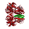
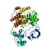
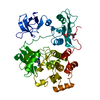
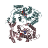
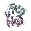
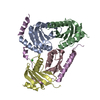
| Title | Crystal Structure of a Periplasmic Heme Binding Protein from Shigella dysenteriae | ||||||
 Components Components | Bacterial heme binding protein | ||||||
 Keywords Keywords | TRANSPORT PROTEIN / periplasmic binding protein / heme transport | ||||||
| Function / homology |  Function and homology information Function and homology information | ||||||
| Biological species |  Shigella dysenteriae (bacteria) Shigella dysenteriae (bacteria) | ||||||
| Method |  X-RAY DIFFRACTION / SYNCHROTRON / MOLECULAR REPLACEMENT / Resolution: 2.05 Å X-RAY DIFFRACTION / SYNCHROTRON / MOLECULAR REPLACEMENT / Resolution: 2.05 Å | ||||||
 Authors Authors | Ho, W.W. / Li, H. / Poulos, T.L. | ||||||
 Citation Citation | Journal: J.Biol.Chem. / Year: 2007 Title: Holo- and apo-bound structures of bacterial periplasmic heme-binding proteins. Authors: Ho, W.W. / Li, H. / Eakanunkul, S. / Tong, Y. / Wilks, A. / Guo, M. / Poulos, T.L. | ||||||
| History |
|
- Structure visualization
| Structure viewer | Molecule: MolmilJmol/JSmol |
|---|
- Downloads & links
-Download
| PDBx/mmCIF format | 2r7a.cif.gz | 212.6 KB | Display | PDBx/mmCIF format |
|---|---|---|---|---|
| PDB format | pdb2r7a.ent.gz | 168.7 KB | Display | PDB format |
| PDBx/mmJSON format | 2r7a.json.gz | Tree view | PDBx/mmJSON format | |
| Others |  Other downloads Other downloads |
-Validation report
| Arichive directory | https://data.pdbj.org/pub/pdb/validation_reports/r7/2r7aftp://data.pdbj.org/pub/pdb/validation_reports/r7/2r7a | HTTPS FTP |
|---|
-Related structure data
| Related structure data | 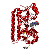 2r79SC 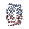 2rg7C S: Starting model for refinement C: citing same article ( |
|---|---|
| Similar structure data |


-Links
 PDBj
PDBj- Assembly
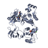
| Deposited unit | 
| ||||||||
|---|---|---|---|---|---|---|---|---|---|
| 1 | 
| ||||||||
| 2 | 
| ||||||||
| Unit cell |
|
-Components
| #1: Protein | Mass: 27582.484 Da / Num. of mol.: 4 Source method: isolated from a genetically manipulated source Source: (gene. exp.) Shigella dysenteriae (bacteria) / Strain: pSHU262 / Plasmid: pSHU262 / Species (production host): Escherichia coli / Production host: #2: Chemical | ChemComp-HEM / |   Mass: 616.487 Da / Num. of mol.: 1 / Source method: obtained synthetically / Formula: C34H32FeN4O4 Mass: 616.487 Da / Num. of mol.: 1 / Source method: obtained synthetically / Formula: C34H32FeN4O4#3: Water | ChemComp-HOH / |  Mass: 18.015 Da / Num. of mol.: 499 / Source method: isolated from a natural source / Formula: H2O Mass: 18.015 Da / Num. of mol.: 499 / Source method: isolated from a natural source / Formula: H2O |
|---|
-Experimental details
-Experiment
| Experiment | Method: X-RAY DIFFRACTION / Number of used crystals: 1 |
|---|
- Sample preparation
| Crystal | Density Matthews: 2.46 Å3/Da / Density % sol: 49.93 % |
|---|---|
| Crystal grow | Temperature: 298 K / Method: vapor diffusion, hanging drop / pH: 4 Details: 0.1M sodium citrate, 0.05M ammonium bromide, 15% polyethylene glycol 4000, 0.001M hemin chloride, pH 4.0, VAPOR DIFFUSION, HANGING DROP, temperature 298K |
-Data collection
| Diffraction | Mean temperature: 100 K |
|---|---|
| Diffraction source | Source: SYNCHROTRON / Site: SSRL  / Beamline: BL9-2 / Wavelength: 1 Å / Beamline: BL9-2 / Wavelength: 1 Å |
| Detector | Type: MARMOSAIC 325 mm CCD / Detector: CCD / Date: Jun 6, 2006 / Details: mirrors |
| Radiation | Monochromator: GRAPHITE / Protocol: SINGLE WAVELENGTH / Monochromatic (M) / Laue (L): M / Scattering type: x-ray |
| Radiation wavelength | Wavelength: 1 Å / Relative weight: 1 |
| Reflection | Resolution: 2.05→43.86 Å / Num. obs: 63384 / % possible obs: 98 % / Observed criterion σ(I): -3 / Redundancy: 3.9 % / Biso Wilson estimate: 19.7 Å2 / Rmerge(I) obs: 0.057 / Rsym value: 0.057 / Net I/σ(I): 27.2 |
| Reflection shell | Resolution: 2.05→2.09 Å / Redundancy: 3.9 % / Mean I/σ(I) obs: 2.01 / Rsym value: 0.67 / % possible all: 97 |
- Processing
| Software |
| ||||||||||||||||||||||||||||||||||||||||||||||||||||||||||||||||||||||||||||||||
|---|---|---|---|---|---|---|---|---|---|---|---|---|---|---|---|---|---|---|---|---|---|---|---|---|---|---|---|---|---|---|---|---|---|---|---|---|---|---|---|---|---|---|---|---|---|---|---|---|---|---|---|---|---|---|---|---|---|---|---|---|---|---|---|---|---|---|---|---|---|---|---|---|---|---|---|---|---|---|---|---|---|
| Refinement | Method to determine structure: MOLECULAR REPLACEMENT Starting model: model of PhuT, PDB ENTRY 2R79 Resolution: 2.05→43.86 Å / Rfactor Rfree error: 0.003 / Data cutoff high absF: 1452602.24 / Data cutoff low absF: 0 / Isotropic thermal model: RESTRAINED / Cross valid method: THROUGHOUT / σ(F): 0 / Stereochemistry target values: Engh & Huber / Details: BULK SOLVENT MODEL USED
| ||||||||||||||||||||||||||||||||||||||||||||||||||||||||||||||||||||||||||||||||
| Solvent computation | Solvent model: FLAT MODEL / Bsol: 66.0413 Å2 / ksol: 0.4 e/Å3 | ||||||||||||||||||||||||||||||||||||||||||||||||||||||||||||||||||||||||||||||||
| Displacement parameters | Biso mean: 39 Å2
| ||||||||||||||||||||||||||||||||||||||||||||||||||||||||||||||||||||||||||||||||
| Refine analyze |
| ||||||||||||||||||||||||||||||||||||||||||||||||||||||||||||||||||||||||||||||||
| Refinement step | Cycle: LAST / Resolution: 2.05→43.86 Å
| ||||||||||||||||||||||||||||||||||||||||||||||||||||||||||||||||||||||||||||||||
| Refine LS restraints |
| ||||||||||||||||||||||||||||||||||||||||||||||||||||||||||||||||||||||||||||||||
| LS refinement shell | Resolution: 2.05→2.18 Å / Rfactor Rfree error: 0.009 / Total num. of bins used: 6
| ||||||||||||||||||||||||||||||||||||||||||||||||||||||||||||||||||||||||||||||||
| Xplor file |
|
