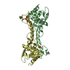
 Movie
Movie Controller


+ Open data

- Basic information
| Entry | Database: PDB / ID: 2r50 | ||||||
|---|---|---|---|---|---|---|---|
| Title | The crystal structure of nonsymbiotic corn hemoglobin 1 | ||||||
 Components Components | Non-symbiotic hemoglobin | ||||||
 Keywords Keywords | METAL BINDING PROTEIN / corn hemoglobin / plant hemoglobin / nonsymbiotic hemoglobin / Heme / Iron / Metal-binding | ||||||
| Function / homology |  Function and homology information Function and homology informationOxidoreductases; Acting on other nitrogenous compounds as donors; With a cytochrome as acceptor / oxygen binding / oxidoreductase activity / heme binding / metal ion binding / nucleus / cytoplasm Similarity search - Function | ||||||
| Biological species |  | ||||||
| Method |  X-RAY DIFFRACTION / MOLECULAR REPLACEMENT / molecular replacement / Resolution: 2.2 Å X-RAY DIFFRACTION / MOLECULAR REPLACEMENT / molecular replacement / Resolution: 2.2 Å | ||||||
 Authors Authors | Smagghe, B.J. / Hoy, J.A. / Hargrove, M.S. | ||||||
 Citation Citation | Journal: To be Published Title: The crystal structure of nonsymbiotic corn hemoglobin 1 Authors: Smagghe, B.J. / Hoy, J.A. / Hargrove, M.S. | ||||||
| History |
|
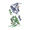
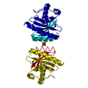
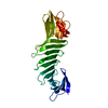
- Structure visualization
| Structure viewer | Molecule: MolmilJmol/JSmol |
|---|
- Downloads & links
-Download
| PDBx/mmCIF format | 2r50.cif.gz | 146.1 KB | Display | PDBx/mmCIF format |
|---|---|---|---|---|
| PDB format | pdb2r50.ent.gz | 114.5 KB | Display | PDB format |
| PDBx/mmJSON format | 2r50.json.gz | Tree view | PDBx/mmJSON format | |
| Others |  Other downloads Other downloads |
-Validation report
| Summary document | 2r50_validation.pdf.gz | 1.8 MB | Display | wwPDB validaton report |
|---|---|---|---|---|
| Full document | 2r50_full_validation.pdf.gz | 1.8 MB | Display | |
| Data in XML | 2r50_validation.xml.gz | 33.9 KB | Display | |
| Data in CIF | 2r50_validation.cif.gz | 47.5 KB | Display | |
| Arichive directory | https://data.pdbj.org/pub/pdb/validation_reports/r5/2r50ftp://data.pdbj.org/pub/pdb/validation_reports/r5/2r50 | HTTPS FTP |
-Related structure data
| Related structure data |  1d8uS S: Starting model for refinement |
|---|---|
| Similar structure data |
-Links
 PDBj
PDBj


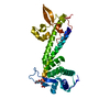
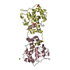
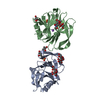
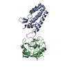
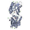
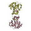
- Assembly
| Deposited unit | 
| ||||||||
|---|---|---|---|---|---|---|---|---|---|
| 1 | 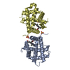
| ||||||||
| 2 | 
| ||||||||
| 3 |
| ||||||||
| Unit cell |
|
-Components
| #1: Protein | Mass: 18304.375 Da / Num. of mol.: 4 Source method: isolated from a genetically manipulated source Source: (gene. exp.) Species: Zea mays / Strain: subsp. parviglumis / Gene: HB, GLB1 / Plasmid: pET28a / Production host:  #2: Chemical | ChemComp-HEM /   Mass: 616.487 Da / Num. of mol.: 4 / Source method: obtained synthetically / Formula: C34H32FeN4O4 Mass: 616.487 Da / Num. of mol.: 4 / Source method: obtained synthetically / Formula: C34H32FeN4O4#3: Chemical | ChemComp-SO4 /   Mass: 96.063 Da / Num. of mol.: 5 / Source method: obtained synthetically / Formula: SO4 Mass: 96.063 Da / Num. of mol.: 5 / Source method: obtained synthetically / Formula: SO4#4: Chemical |   Mass: 60.052 Da / Num. of mol.: 2 / Source method: obtained synthetically / Formula: C2H4O2 Mass: 60.052 Da / Num. of mol.: 2 / Source method: obtained synthetically / Formula: C2H4O2#5: Water | ChemComp-HOH / |  Mass: 18.015 Da / Num. of mol.: 584 / Source method: isolated from a natural source / Formula: H2O Mass: 18.015 Da / Num. of mol.: 584 / Source method: isolated from a natural source / Formula: H2O |
|---|
-Experimental details
-Experiment
| Experiment | Method: X-RAY DIFFRACTION / Number of used crystals: 1 |
|---|
- Sample preparation
| Crystal | Density Matthews: 2.45 Å3/Da / Density % sol: 49.76 % |
|---|---|
| Crystal grow | Temperature: 277 K / Method: vapor diffusion, hanging drop / pH: 5.6 Details: 0.1M Sodium Acetate, 0.2M Ammonium Sulfate, 20% PEG MME 1900, pH 5.6, VAPOR DIFFUSION, HANGING DROP, temperature 277K |
-Data collection
| Diffraction | Mean temperature: 100 K |
|---|---|
| Diffraction source | Source: ROTATING ANODE / Type: RIGAKU RU200 / Wavelength: 1.54 Å |
| Detector | Type: RIGAKU RAXIS IV / Detector: IMAGE PLATE / Date: Jan 23, 2004 / Details: Osmic |
| Radiation | Protocol: SINGLE WAVELENGTH / Monochromatic (M) / Laue (L): M / Scattering type: x-ray |
| Radiation wavelength | Wavelength: 1.54 Å / Relative weight: 1 |
| Reflection | Resolution: 2.2→80 Å / Num. obs: 35371 / % possible obs: 99.6 % / Redundancy: 6.67 % / Biso Wilson estimate: 38.8 Å2 / Rmerge(I) obs: 0.05 / Net I/σ(I): 19.3 |
| Reflection shell | Resolution: 2.2→2.26 Å / Redundancy: 5.24 % / Rmerge(I) obs: 0.24 / Mean I/σ(I) obs: 6.4 / Num. unique all: 2504 / % possible all: 97.6 |
-Phasing
| Phasing | Method: molecular replacement |
|---|
- Processing
| Software |
| ||||||||||||||||||||||||||||||||||||||||||||||||||||||||||||||||||||||||||||||||||||||||||||||||||||
|---|---|---|---|---|---|---|---|---|---|---|---|---|---|---|---|---|---|---|---|---|---|---|---|---|---|---|---|---|---|---|---|---|---|---|---|---|---|---|---|---|---|---|---|---|---|---|---|---|---|---|---|---|---|---|---|---|---|---|---|---|---|---|---|---|---|---|---|---|---|---|---|---|---|---|---|---|---|---|---|---|---|---|---|---|---|---|---|---|---|---|---|---|---|---|---|---|---|---|---|---|---|
| Refinement | Method to determine structure: MOLECULAR REPLACEMENT Starting model: pdb entry 1D8U Resolution: 2.2→79.06 Å / Cor.coef. Fo:Fc: 0.952 / Cor.coef. Fo:Fc free: 0.922 / SU B: 6.762 / SU ML: 0.166 / Cross valid method: THROUGHOUT / σ(F): 0 / ESU R: 0.28 / ESU R Free: 0.221 / Stereochemistry target values: MAXIMUM LIKELIHOOD / Details: HYDROGENS HAVE BEEN ADDED IN THE RIDING POSITIONS
| ||||||||||||||||||||||||||||||||||||||||||||||||||||||||||||||||||||||||||||||||||||||||||||||||||||
| Solvent computation | Ion probe radii: 0.8 Å / Shrinkage radii: 0.8 Å / VDW probe radii: 1.4 Å / Solvent model: BABINET MODEL WITH MASK | ||||||||||||||||||||||||||||||||||||||||||||||||||||||||||||||||||||||||||||||||||||||||||||||||||||
| Displacement parameters | Biso mean: 38.837 Å2
| ||||||||||||||||||||||||||||||||||||||||||||||||||||||||||||||||||||||||||||||||||||||||||||||||||||
| Refinement step | Cycle: LAST / Resolution: 2.2→79.06 Å
| ||||||||||||||||||||||||||||||||||||||||||||||||||||||||||||||||||||||||||||||||||||||||||||||||||||
| Refine LS restraints |
| ||||||||||||||||||||||||||||||||||||||||||||||||||||||||||||||||||||||||||||||||||||||||||||||||||||
| LS refinement shell | Resolution: 2.2→2.257 Å / Total num. of bins used: 20
|
