 Movie
Movie Controller


+ Open data

- Basic information
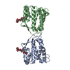
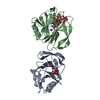
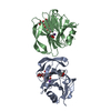
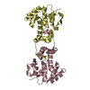
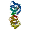
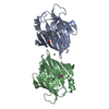
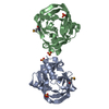
| Entry | Database: PDB / ID: 1aih | ||||||
|---|---|---|---|---|---|---|---|
| Title | CATALYTIC DOMAIN OF BACTERIOPHAGE HP1 INTEGRASE | ||||||
 Components Components | HP1 INTEGRASE | ||||||
 Keywords Keywords | DNA INTEGRATION / RECOMBINATION | ||||||
| Function / homology |  Function and homology information Function and homology informationDNA integration / viral genome integration into host DNA / establishment of integrated proviral latency / Transferases; Transferring phosphorus-containing groups; Nucleotidyltransferases / transferase activity / DNA recombination / Hydrolases; Acting on ester bonds / hydrolase activity / symbiont entry into host cell / DNA binding Similarity search - Function | ||||||
| Biological species |  Haemophilus phage HP1 (virus) Haemophilus phage HP1 (virus) | ||||||
| Method |  X-RAY DIFFRACTION / SYNCHROTRON / MAD / Resolution: 2.5 Å X-RAY DIFFRACTION / SYNCHROTRON / MAD / Resolution: 2.5 Å | ||||||
 Authors Authors | Hickman, A.B. / Waninger, S. / Scocca, J.J. / Dyda, F. | ||||||
 Citation Citation | Journal: Cell(Cambridge,Mass.) / Year: 1997 Title: Molecular organization in site-specific recombination: the catalytic domain of bacteriophage HP1 integrase at 2.7 A resolution. Authors: Hickman, A.B. / Waninger, S. / Scocca, J.J. / Dyda, F. | ||||||
| History |
|
- Structure visualization
| Structure viewer | Molecule: MolmilJmol/JSmol |
|---|
- Downloads & links
-Download
| PDBx/mmCIF format | 1aih.cif.gz | 147.6 KB | Display | PDBx/mmCIF format |
|---|---|---|---|---|
| PDB format | pdb1aih.ent.gz | 116.9 KB | Display | PDB format |
| PDBx/mmJSON format | 1aih.json.gz | Tree view | PDBx/mmJSON format | |
| Others |  Other downloads Other downloads |
-Validation report
| Arichive directory | https://data.pdbj.org/pub/pdb/validation_reports/ai/1aihftp://data.pdbj.org/pub/pdb/validation_reports/ai/1aih | HTTPS FTP |
|---|
-Related structure data
| Similar structure data |
|---|


-Links
 PDBj
PDBj


- Assembly
| Deposited unit | 
| ||||||||||||
|---|---|---|---|---|---|---|---|---|---|---|---|---|---|
| 1 | 
| ||||||||||||
| 2 | 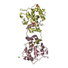
| ||||||||||||
| Unit cell |
| ||||||||||||
| Noncrystallographic symmetry (NCS) | NCS oper:
|
-Components
| #1: Protein | Mass: 19457.312 Da / Num. of mol.: 4 / Fragment: CATALYTIC DOMAIN, RESIDUES 168 - 337 Source method: isolated from a genetically manipulated source Source: (gene. exp.) Haemophilus phage HP1 (virus) / Genus: P2-like viruses / Strain: HP1C1 / Cell line: HAEMOPHILUS INFLUENZAE L10 / Gene: GENBANK U24159 / Plasmid: PHPC / Production host:  #2: Chemical | ChemComp-SO4 /   Mass: 96.063 Da / Num. of mol.: 8 / Source method: obtained synthetically / Formula: SO4 Mass: 96.063 Da / Num. of mol.: 8 / Source method: obtained synthetically / Formula: SO4#3: Chemical | ChemComp-MG /   Mass: 24.305 Da / Num. of mol.: 5 / Source method: obtained synthetically / Formula: Mg Mass: 24.305 Da / Num. of mol.: 5 / Source method: obtained synthetically / Formula: Mg#4: Water | ChemComp-HOH / |  Mass: 18.015 Da / Num. of mol.: 152 / Source method: isolated from a natural source / Formula: H2O Mass: 18.015 Da / Num. of mol.: 152 / Source method: isolated from a natural source / Formula: H2O |
|---|
-Experimental details
-Experiment
| Experiment | Method: X-RAY DIFFRACTION / Number of used crystals: 2 |
|---|
- Sample preparation
| Crystal | Density Matthews: 3.98 Å3/Da / Density % sol: 69 % | ||||||||||||||||||||||||||||||
|---|---|---|---|---|---|---|---|---|---|---|---|---|---|---|---|---|---|---|---|---|---|---|---|---|---|---|---|---|---|---|---|
| Crystal grow | Method: vapor diffusion / pH: 7.5 Details: VAPOR DIFFUSION, 15% PEG 8000, 45MM NA CAODYLATE, 34MM AMMONIUM SULFATE, 10MM TRIS-HCL, 4.5% GLYCEROL, 0.1 M MGCL., pH 7.5, vapor diffusion | ||||||||||||||||||||||||||||||
| Crystal grow | *PLUS pH: 6.5 / Method: vapor diffusion, hanging drop | ||||||||||||||||||||||||||||||
| Components of the solutions | *PLUS
|
-Data collection
| Diffraction | Mean temperature: 95 K |
|---|---|
| Diffraction source | Source: SYNCHROTRON / Site: CHESS  / Beamline: F2 / Wavelength: 1.6498 / Beamline: F2 / Wavelength: 1.6498 |
| Detector | Type: PRINCETON 2K / Detector: CCD / Date: Dec 12, 1996 / Details: BENT MIRROR |
| Radiation | Monochromator: SI(111) / Monochromatic (M) / Laue (L): M / Scattering type: x-ray |
| Radiation wavelength | Wavelength: 1.6498 Å / Relative weight: 1 |
| Reflection | Resolution: 2.7→30 Å / Num. obs: 34819 / % possible obs: 97 % / Observed criterion σ(I): 0 / Redundancy: 6 % / Biso Wilson estimate: 42 Å2 / Rmerge(I) obs: 0.087 / Rsym value: 0.087 / Net I/σ(I): 11.4 |
| Reflection shell | Resolution: 2.7→2.78 Å / Redundancy: 3 % / Rmerge(I) obs: 0.18 / Mean I/σ(I) obs: 5 / Rsym value: 0.18 / % possible all: 89.7 |
| Reflection | *PLUS Num. measured all: 52126 |
| Reflection shell | *PLUS % possible obs: 89.7 % |
- Processing
| Software |
| ||||||||||||||||||||||||||||||||||||||||||||||||||||||||||||
|---|---|---|---|---|---|---|---|---|---|---|---|---|---|---|---|---|---|---|---|---|---|---|---|---|---|---|---|---|---|---|---|---|---|---|---|---|---|---|---|---|---|---|---|---|---|---|---|---|---|---|---|---|---|---|---|---|---|---|---|---|---|
| Refinement | Method to determine structure: MAD / Resolution: 2.5→30 Å / Rfactor Rfree error: 0.006 / Data cutoff high absF: 100000 / Data cutoff low absF: 0.1 / Cross valid method: THROUGHOUT / σ(F): 2
| ||||||||||||||||||||||||||||||||||||||||||||||||||||||||||||
| Displacement parameters | Biso mean: 47.77 Å2 | ||||||||||||||||||||||||||||||||||||||||||||||||||||||||||||
| Refine analyze | Luzzati d res low obs: 30 Å | ||||||||||||||||||||||||||||||||||||||||||||||||||||||||||||
| Refinement step | Cycle: LAST / Resolution: 2.5→30 Å
| ||||||||||||||||||||||||||||||||||||||||||||||||||||||||||||
| Refine LS restraints |
| ||||||||||||||||||||||||||||||||||||||||||||||||||||||||||||
| Refine LS restraints NCS | NCS model details: UNRESTRAINED | ||||||||||||||||||||||||||||||||||||||||||||||||||||||||||||
| LS refinement shell | Resolution: 2.5→2.61 Å / Rfactor Rfree error: 0.04 / Total num. of bins used: 8
| ||||||||||||||||||||||||||||||||||||||||||||||||||||||||||||
| Xplor file |
| ||||||||||||||||||||||||||||||||||||||||||||||||||||||||||||
| Software | *PLUS Name: X-PLOR / Version: 3.1 / Classification: refinement | ||||||||||||||||||||||||||||||||||||||||||||||||||||||||||||
| Refinement | *PLUS | ||||||||||||||||||||||||||||||||||||||||||||||||||||||||||||
| Solvent computation | *PLUS | ||||||||||||||||||||||||||||||||||||||||||||||||||||||||||||
| Displacement parameters | *PLUS | ||||||||||||||||||||||||||||||||||||||||||||||||||||||||||||
| Refine LS restraints | *PLUS
| ||||||||||||||||||||||||||||||||||||||||||||||||||||||||||||
| LS refinement shell | *PLUS Rfactor obs: 0.318 |
