Type: MAR CCD 165 mm / Detector: CCD / Date: Jun 28, 2005 / Details: mirrors
Radiation
Monochromator: GRAPHITE / Protocol: SINGLE WAVELENGTH / Monochromatic (M) / Laue (L): M / Scattering type: x-ray
Radiation wavelength
Wavelength: 0.954 Å / Relative weight: 1
Reflection
Av σ(I) over netI: 14.7 / Number: 21402 / Rmerge(I) obs: 0.075 / Χ2: 1.03 / D res high: 2.5 Å / D res low: 30 Å / Num. obs: 6704 / % possible obs: 94.6
Diffraction reflection shell
Highest resolution (Å)
Lowest resolution (Å)
% possible obs (%)
ID
Rmerge(I) obs
Chi squared
5.38
30
98.8
1
0.054
0.752
4.27
5.38
99.9
1
0.061
0.765
3.73
4.27
100
1
0.068
1.058
3.39
3.73
99.7
1
0.08
1.058
3.15
3.39
99.9
1
0.091
1.057
2.96
3.15
99.9
1
0.11
0.981
2.82
2.96
98.9
1
0.125
1.155
2.69
2.82
92.7
1
0.13
1.201
2.59
2.69
83.7
1
0.132
1.355
2.5
2.59
71.6
1
0.118
1.433
Reflection
Resolution: 2.5→30 Å / Num. obs: 6704 / % possible obs: 94.6 % / Redundancy: 3.2 % / Biso Wilson estimate: 43.8 Å2 / Rmerge(I) obs: 0.075 / Χ2: 1.033 / Net I/σ(I): 14.7
Reflection shell
Resolution (Å)
Rmerge(I) obs
Num. unique all
Χ2
% possible all
2.5-2.59
0.118
486
1.433
71.6
2.59-2.69
0.132
605
1.355
83.7
2.69-2.82
0.13
650
1.201
92.7
2.82-2.96
0.125
692
1.155
98.9
2.96-3.15
0.11
702
0.981
99.9
3.15-3.39
0.091
712
1.057
99.9
3.39-3.73
0.08
711
1.058
99.7
3.73-4.27
0.068
708
1.058
100
4.27-5.38
0.061
711
0.765
99.9
5.38-30
0.054
727
0.752
98.8
-
Phasing
Phasing
Method: molecular replacement
Phasing MR
Highest resolution
Lowest resolution
Rotation
3 Å
25.08 Å
Translation
3 Å
25.08 Å
-
Processing
Software
Name
Version
Classification
NB
DENZO
datareduction
SCALEPACK
datascaling
MOLREP
phasing
REFMAC
5.2.0019
refinement
PDB_EXTRACT
3
dataextraction
MAR345dtb
datacollection
Refinement
Method to determine structure: MOLECULAR REPLACEMENT / Resolution: 2.5→25.07 Å / Cor.coef. Fo:Fc: 0.923 / Cor.coef. Fo:Fc free: 0.854 / SU B: 10.53 / SU ML: 0.23 / Cross valid method: THROUGHOUT / σ(F): 0 / ESU R: 0.598 / ESU R Free: 0.338 / Stereochemistry target values: MAXIMUM LIKELIHOOD / Details: HYDROGENS HAVE BEEN ADDED IN THE RIDING POSITIONS
Rfactor
Num. reflection
% reflection
Selection details
Rfree
0.287
313
4.7 %
RANDOM
Rwork
0.21
-
-
-
obs
0.214
6693
94.6 %
-
Solvent computation
Ion probe radii: 0.8 Å / Shrinkage radii: 0.8 Å / VDW probe radii: 1.4 Å / Solvent model: MASK
Displacement parameters
Biso mean: 27.726 Å2
Baniso -1
Baniso -2
Baniso -3
1-
-1.44 Å2
0 Å2
0.69 Å2
2-
-
0.07 Å2
0 Å2
3-
-
-
1.94 Å2
Refinement step
Cycle: LAST / Resolution: 2.5→25.07 Å
Protein
Nucleic acid
Ligand
Solvent
Total
Num. atoms
1220
0
5
105
1330
Refine LS restraints
Refine-ID
Type
Dev ideal
Dev ideal target
Number
X-RAY DIFFRACTION
r_bond_refined_d
0.009
0.022
1243
X-RAY DIFFRACTION
r_angle_refined_deg
1.197
2.01
1685
X-RAY DIFFRACTION
r_dihedral_angle_1_deg
5.101
5
153
X-RAY DIFFRACTION
r_dihedral_angle_2_deg
37.878
26.667
54
X-RAY DIFFRACTION
r_dihedral_angle_3_deg
17.182
15
224
X-RAY DIFFRACTION
r_dihedral_angle_4_deg
6.7
15
2
X-RAY DIFFRACTION
r_chiral_restr
0.085
0.2
189
X-RAY DIFFRACTION
r_gen_planes_refined
0.003
0.02
918
X-RAY DIFFRACTION
r_nbd_refined
0.218
0.2
620
X-RAY DIFFRACTION
r_nbtor_refined
0.301
0.2
864
X-RAY DIFFRACTION
r_xyhbond_nbd_refined
0.12
0.2
74
X-RAY DIFFRACTION
r_symmetry_vdw_refined
0.178
0.2
38
X-RAY DIFFRACTION
r_symmetry_hbond_refined
0.122
0.2
6
X-RAY DIFFRACTION
r_mcbond_it
0.458
1.5
811
X-RAY DIFFRACTION
r_mcangle_it
0.791
2
1255
X-RAY DIFFRACTION
r_scbond_it
0.912
3
496
X-RAY DIFFRACTION
r_scangle_it
1.474
4.5
430
LS refinement shell
Resolution: 2.5→2.565 Å / Total num. of bins used: 20
Rfactor
Num. reflection
% reflection
Rfree
0.329
13
-
Rwork
0.264
326
-
all
-
339
-
obs
-
-
68.48 %
+
About Yorodumi
-
News
-
Feb 9, 2022. New format data for meta-information of EMDB entries
New format data for meta-information of EMDB entries
Version 3 of the EMDB header file is now the official format.
The previous official version 1.9 will be removed from the archive.
In the structure databanks used in Yorodumi, some data are registered as the other names, "COVID-19 virus" and "2019-nCoV". Here are the details of the virus and the list of structure data.
Jan 31, 2019. EMDB accession codes are about to change! (news from PDBe EMDB page)
EMDB accession codes are about to change! (news from PDBe EMDB page)
The allocation of 4 digits for EMDB accession codes will soon come to an end. Whilst these codes will remain in use, new EMDB accession codes will include an additional digit and will expand incrementally as the available range of codes is exhausted. The current 4-digit format prefixed with “EMD-” (i.e. EMD-XXXX) will advance to a 5-digit format (i.e. EMD-XXXXX), and so on. It is currently estimated that the 4-digit codes will be depleted around Spring 2019, at which point the 5-digit format will come into force.
The EM Navigator/Yorodumi systems omit the EMD- prefix.
Related info.:Q: What is EMD? / ID/Accession-code notation in Yorodumi/EM Navigator
Yorodumi is a browser for structure data from EMDB, PDB, SASBDB, etc.
This page is also the successor to EM Navigator detail page, and also detail information page/front-end page for Omokage search.
The word "yorodu" (or yorozu) is an old Japanese word meaning "ten thousand". "mi" (miru) is to see.
Related info.:EMDB / PDB / SASBDB / Comparison of 3 databanks / Yorodumi Search / Aug 31, 2016. New EM Navigator & Yorodumi / Yorodumi Papers / Jmol/JSmol / Function and homology information / Changes in new EM Navigator and Yorodumi
 Movie
Movie Controller


 Open data
 Basic information
 Components
 Keywords
 Function and homology information
 Homo sapiens (human)
 X-RAY DIFFRACTION /
  Authors
 Citation
 Structure visualization
 Downloads & links
 Other downloads


 PDBj
PDBj


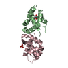
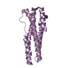
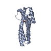
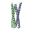
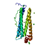
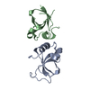
 Assembly
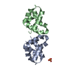
 Pichia pastoris (fungus) / Strain (production host): GS115 / References: UniProt: P07602
 Mass: 96.063 Da / Num. of mol.: 1 / Source method: obtained synthetically / Formula: SO4
 Mass: 18.015 Da / Num. of mol.: 105 / Source method: isolated from a natural source / Formula: H2O
 Sample preparation
 / Beamline: 14.1 / Wavelength: 0.954 Å
 Processing