 Movie
Movie Controller

[English] 日本語
 Yorodumi
- PDB-1mu4: CRYSTAL STRUCTURE AT 1.8 ANGSTROMS OF THE BACILLUS SUBTILIS CATAB... -
+ Open data

- Basic information
| Entry | Database: PDB / ID: 1mu4 | ||||||
|---|---|---|---|---|---|---|---|
| Title | CRYSTAL STRUCTURE AT 1.8 ANGSTROMS OF THE BACILLUS SUBTILIS CATABOLITE REPRESSION HISTIDINE CONTAINING PROTEIN (CRH) | ||||||
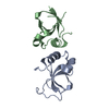
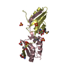
 Components Components | HPr-like protein crh | ||||||
 Keywords Keywords | TRANSPORT PROTEIN / OPEN-FACED B-SANDWICH / PHOSPHOTRANSFERASE SYSTEM / SWAPPING DOMAIN | ||||||
| Function / homology |  Function and homology information Function and homology information | ||||||
| Biological species |  | ||||||
| Method |  X-RAY DIFFRACTION / SYNCHROTRON / MOLECULAR REPLACEMENT / Resolution: 1.8 Å X-RAY DIFFRACTION / SYNCHROTRON / MOLECULAR REPLACEMENT / Resolution: 1.8 Å | ||||||
 Authors Authors | Juy, M.R. / Haser, R. | ||||||
 Citation Citation | Journal: J.Mol.Biol. / Year: 2003 Title: Dimerization of Crh by reversible 3D Domain Swapping Induces Structural Adjustments to its monomeric homologue HPR Authors: Juy, M.R. / Penin, F. / Favier, A. / Galinier, A. / Montserret, R. / Haser, R. / Deutscher, J. / Bockmann, A. #1: Journal: J.MOL.MICROBIOL.BIOTECHNOL. / Year: 2001Title: Evidence for a Dimerisation State of the Bacillus Subtilis Catabolite Repression Hpr-Like Protein, Crh Authors: Penin, F. / Favier, A. / Montserret, R. / Brutscher, B. / Deutscher, J. / Marion, D. / Galinier, D. | ||||||
| History |
|
- Structure visualization
| Structure viewer | Molecule: MolmilJmol/JSmol |
|---|
- Downloads & links
-Download
| PDBx/mmCIF format | 1mu4.cif.gz | 46.5 KB | Display | PDBx/mmCIF format |
|---|---|---|---|---|
| PDB format | pdb1mu4.ent.gz | 34.2 KB | Display | PDB format |
| PDBx/mmJSON format | 1mu4.json.gz | Tree view | PDBx/mmJSON format | |
| Others |  Other downloads Other downloads |
-Validation report
| Arichive directory | https://data.pdbj.org/pub/pdb/validation_reports/mu/1mu4ftp://data.pdbj.org/pub/pdb/validation_reports/mu/1mu4 | HTTPS FTP |
|---|
-Related structure data
| Related structure data | 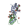 1mo1S S: Starting model for refinement |
|---|---|
| Similar structure data |


-Links
 PDBj
PDBj- Assembly
| Deposited unit | 
| ||||||||
|---|---|---|---|---|---|---|---|---|---|
| 1 |
| ||||||||
| 2 | 
| ||||||||
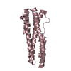
| Unit cell |
|
-Components
| #1: Protein | Mass: 9577.953 Da / Num. of mol.: 2 Source method: isolated from a genetically manipulated source Source: (gene. exp.) #2: Chemical |   Mass: 96.063 Da / Num. of mol.: 3 / Source method: obtained synthetically / Formula: SO4 Mass: 96.063 Da / Num. of mol.: 3 / Source method: obtained synthetically / Formula: SO4#3: Water | ChemComp-HOH / |  Mass: 18.015 Da / Num. of mol.: 122 / Source method: isolated from a natural source / Formula: H2O Mass: 18.015 Da / Num. of mol.: 122 / Source method: isolated from a natural source / Formula: H2O |
|---|
-Experimental details
-Experiment
| Experiment | Method: X-RAY DIFFRACTION / Number of used crystals: 1 |
|---|
- Sample preparation
| Crystal | Density Matthews: 3.71 Å3/Da / Density % sol: 66.82 % | ||||||||||||||||||||||||||||||||||||||||||
|---|---|---|---|---|---|---|---|---|---|---|---|---|---|---|---|---|---|---|---|---|---|---|---|---|---|---|---|---|---|---|---|---|---|---|---|---|---|---|---|---|---|---|---|
| Crystal grow | Temperature: 297 K / Method: vapor diffusion, hanging drop / pH: 6.5 Details: ammonium sulfate, peg 1000,, pH 6.5, VAPOR DIFFUSION, HANGING DROP, temperature 297K | ||||||||||||||||||||||||||||||||||||||||||
| Crystal grow | *PLUS Temperature: 18 ℃ / Method: vapor diffusion, hanging drop | ||||||||||||||||||||||||||||||||||||||||||
| Components of the solutions | *PLUS
|
-Data collection
| Diffraction | Mean temperature: 297 K |
|---|---|
| Diffraction source | Source: SYNCHROTRON / Site: ESRF  / Type: ESRF / Wavelength: 0.9 / Type: ESRF / Wavelength: 0.9 |
| Detector | Type: MARRESEARCH / Detector: IMAGE PLATE / Date: May 8, 2001 |
| Radiation | Monochromator: SI 111 CHANNEL / Protocol: SINGLE WAVELENGTH / Monochromatic (M) / Laue (L): M / Scattering type: x-ray |
| Radiation wavelength | Wavelength: 0.9 Å / Relative weight: 1 |
| Reflection | Resolution: 1.8→29.85 Å / Num. all: 27502 / Num. obs: 27502 / % possible obs: 99.6 % / Observed criterion σ(F): 0 / Observed criterion σ(I): -3 / Redundancy: 4.1 % / Biso Wilson estimate: 22 Å2 / Rmerge(I) obs: 0.055 / Rsym value: 0.055 / Net I/σ(I): 8.5 |
| Reflection shell | Resolution: 1.8→1.86 Å / Redundancy: 3.4 % / Rmerge(I) obs: 0.318 / Mean I/σ(I) obs: 1.9 / Num. unique all: 2112 / Rsym value: 0.318 / % possible all: 99.3 |
| Reflection | *PLUS Highest resolution: 1.8 Å / Lowest resolution: 30 Å / Num. obs: 27478 |
| Reflection shell | *PLUS % possible obs: 99.6 % |
- Processing
| Software |
| ||||||||||||||||||||||||||||||||||||
|---|---|---|---|---|---|---|---|---|---|---|---|---|---|---|---|---|---|---|---|---|---|---|---|---|---|---|---|---|---|---|---|---|---|---|---|---|---|
| Refinement | Method to determine structure: MOLECULAR REPLACEMENT Starting model: pdb entry 1MO1 Resolution: 1.8→29.85 Å / Rfactor Rfree error: 0.005 / Data cutoff high absF: 1664964.41 / Data cutoff high rms absF: 1664964.41 / Data cutoff low absF: 0 / Isotropic thermal model: RESTRAINED / Cross valid method: THROUGHOUT / σ(F): 0 / σ(I): -3 / Stereochemistry target values: Engh & Huber
| ||||||||||||||||||||||||||||||||||||
| Solvent computation | Solvent model: FLAT MODEL / Bsol: 33.8 Å2 / ksol: 0.32 e/Å3 | ||||||||||||||||||||||||||||||||||||
| Displacement parameters | Biso mean: 31.5 Å2
| ||||||||||||||||||||||||||||||||||||
| Refine analyze |
| ||||||||||||||||||||||||||||||||||||
| Refinement step | Cycle: LAST / Resolution: 1.8→29.85 Å
| ||||||||||||||||||||||||||||||||||||
| Refine LS restraints |
| ||||||||||||||||||||||||||||||||||||
| LS refinement shell | Resolution: 1.8→1.91 Å / Rfactor Rfree error: 0.016 / Total num. of bins used: 6
| ||||||||||||||||||||||||||||||||||||
| Refinement | *PLUS Lowest resolution: 30 Å / Rfactor Rfree: 0.199 | ||||||||||||||||||||||||||||||||||||
| Solvent computation | *PLUS | ||||||||||||||||||||||||||||||||||||
| Displacement parameters | *PLUS | ||||||||||||||||||||||||||||||||||||
| Refine LS restraints | *PLUS
|
