Movie
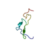
Movie Controller


+ Open data

- Basic information
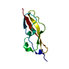
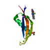
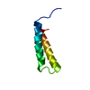
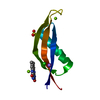
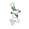
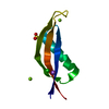
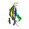
| Entry | Database: PDB / ID: 2qzd | ||||||
|---|---|---|---|---|---|---|---|
| Title | Fitted structure of SCR4 of DAF into cryoEM density | ||||||
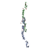
 Components Components | Complement decay-accelerating factor | ||||||
 Keywords Keywords | IMMUNE SYSTEM / SCR4 of DAF from 1ojv fitted into cryoEM density / Blood group antigen / Complement pathway / Glycoprotein / GPI-anchor / Immune response / Innate immunity / Lipoprotein / Membrane / Sushi | ||||||
| Function / homology |  Function and homology information Function and homology informationregulation of lipopolysaccharide-mediated signaling pathway / negative regulation of complement activation / regulation of complement-dependent cytotoxicity / regulation of complement activation / respiratory burst / positive regulation of CD4-positive, alpha-beta T cell activation / positive regulation of CD4-positive, alpha-beta T cell proliferation / Class B/2 (Secretin family receptors) / ficolin-1-rich granule membrane / complement activation, classical pathway ...regulation of lipopolysaccharide-mediated signaling pathway / negative regulation of complement activation / regulation of complement-dependent cytotoxicity / regulation of complement activation / respiratory burst / positive regulation of CD4-positive, alpha-beta T cell activation / positive regulation of CD4-positive, alpha-beta T cell proliferation / Class B/2 (Secretin family receptors) / ficolin-1-rich granule membrane / complement activation, classical pathway / transport vesicle / side of membrane / COPI-mediated anterograde transport / endoplasmic reticulum-Golgi intermediate compartment membrane / secretory granule membrane / Regulation of Complement cascade / positive regulation of T cell cytokine production / positive regulation of cytosolic calcium ion concentration / virus receptor activity / membrane raft / Golgi membrane / innate immune response / lipid binding / Neutrophil degranulation / cell surface / extracellular exosome / extracellular region / plasma membrane Similarity search - Function | ||||||
| Biological species |  Homo sapiens (human) Homo sapiens (human) | ||||||
| Method | ELECTRON MICROSCOPY / single particle reconstruction / cryo EM / Resolution: 14 Å | ||||||
 Authors Authors | Hafenstein, S. / Bowman, V.D. / Chipman, P.R. / Bator Kelly, C.M. / Lin, F. / Medof, M.E. / Rossmann, M.G. | ||||||
 Citation Citation | Journal: J Virol / Year: 2007 Title: Interaction of decay-accelerating factor with coxsackievirus B3. Authors: Susan Hafenstein / Valorie D Bowman / Paul R Chipman / Carol M Bator Kelly / Feng Lin / M Edward Medof / Michael G Rossmann /  Abstract: Many entero-, parecho-, and rhinoviruses use immunoglobulin (Ig)-like receptors that bind into the viral canyon and are required to initiate viral uncoating during infection. However, some of these ...Many entero-, parecho-, and rhinoviruses use immunoglobulin (Ig)-like receptors that bind into the viral canyon and are required to initiate viral uncoating during infection. However, some of these viruses use an alternative or additional receptor that binds outside the canyon. Both the coxsackievirus-adenovirus receptor (CAR), an Ig-like molecule that binds into the viral canyon, and decay-accelerating factor (DAF) have been identified as cellular receptors for coxsackievirus B3 (CVB3). A cryoelectron microscopy reconstruction of a variant of CVB3 complexed with DAF shows full occupancy of the DAF receptor in each of 60 binding sites. The DAF molecule bridges the canyon, blocking the CAR binding site and causing the two receptors to compete with one another. The binding site of DAF on CVB3 differs from the binding site of DAF on the surface of echoviruses, suggesting independent evolutionary processes. | ||||||
| History |
|
- Structure visualization
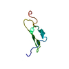
| Movie |
Movie viewer |
|---|---|
| Structure viewer | Molecule: MolmilJmol/JSmol |
 UCSF Chimera
- Downloads & links
-Download
| PDBx/mmCIF format | 2qzd.cif.gz | 24.2 KB | Display | PDBx/mmCIF format |
|---|---|---|---|---|
| PDB format | pdb2qzd.ent.gz | 13.8 KB | Display | PDB format |
| PDBx/mmJSON format | 2qzd.json.gz | Tree view | PDBx/mmJSON format | |
| Others |  Other downloads Other downloads |
-Validation report
| Summary document | 2qzd_validation.pdf.gz | 780 KB | Display | wwPDB validaton report |
|---|---|---|---|---|
| Full document | 2qzd_full_validation.pdf.gz | 779.6 KB | Display | |
| Data in XML | 2qzd_validation.xml.gz | 10 KB | Display | |
| Data in CIF | 2qzd_validation.cif.gz | 12.8 KB | Display | |
| Arichive directory | https://data.pdbj.org/pub/pdb/validation_reports/qz/2qzdftp://data.pdbj.org/pub/pdb/validation_reports/qz/2qzd | HTTPS FTP |
-Related structure data
| Related structure data | 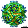 1412MC 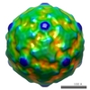 1411C 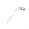 2qzfC 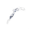 2qzhC M: map data used to model this data C: citing same article ( |
|---|---|
| Similar structure data |
-Links
 PDBj
PDBj


- Assembly
| Deposited unit | 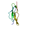
|
|---|---|
| 1 |
|
-Components
| #1: Protein | Mass: 7274.988 Da / Num. of mol.: 1 / Fragment: SCR4 domain Source method: isolated from a genetically manipulated source Source: (gene. exp.) Homo sapiens (human) / Gene: CD55, CR, DAF / Production host:  |
|---|---|
| Has protein modification | Y |
-Experimental details
-Experiment
| Experiment | Method: ELECTRON MICROSCOPY |
|---|---|
| EM experiment | Aggregation state: PARTICLE / 3D reconstruction method: single particle reconstruction |
- Sample preparation
| Component |
| |||||||||||||||
|---|---|---|---|---|---|---|---|---|---|---|---|---|---|---|---|---|
| Buffer solution | Name: 50mM MES / pH: 6 / Details: 50mM MES | |||||||||||||||
| Specimen | Conc.: 2 mg/ml / Embedding applied: NO / Shadowing applied: NO / Staining applied: NO / Vitrification applied: YES | |||||||||||||||
| Specimen support | Details: quantifoils | |||||||||||||||
| Vitrification | Instrument: HOMEMADE PLUNGER / Cryogen name: ETHANE / Details: blot before plunging |
- Electron microscopy imaging
| Microscopy | Model: FEI/PHILIPS CM300FEG/T / Date: Aug 6, 2004 |
|---|---|
| Electron gun | Electron source: TUNGSTEN HAIRPIN / Accelerating voltage: 300 kV / Illumination mode: FLOOD BEAM |
| Electron lens | Mode: BRIGHT FIELD / Nominal magnification: 45000 X / Nominal defocus max: 4.6 nm / Nominal defocus min: 1 nm / Cs: 2 mm |
| Specimen holder | Temperature: 93 K / Tilt angle min: 0 ° |
| Image recording | Electron dose: 24 e/Å2 / Film or detector model: KODAK SO-163 FILM |
| Radiation | Protocol: SINGLE WAVELENGTH / Monochromatic (M) / Laue (L): M / Scattering type: x-ray |
| Radiation wavelength | Relative weight: 1 |
- Processing
| EM software |
| ||||||||||||
|---|---|---|---|---|---|---|---|---|---|---|---|---|---|
| Symmetry | Point symmetry: I (icosahedral) | ||||||||||||
| 3D reconstruction | Resolution: 14 Å / Num. of particles: 2269 / Symmetry type: POINT | ||||||||||||
| Atomic model building | PDB-ID: 1OJV Accession code: 1OJV / Source name: PDB / Type: experimental model | ||||||||||||
| Refinement step | Cycle: LAST
|