 Movie
Movie Controller

[English] 日本語
 Yorodumi
- PDB-2qxi: High resolution structure of Human Kallikrein 7 in Complex with S... -
+ Open data

- Basic information
| Entry | Database: PDB / ID: 2qxi | ||||||
|---|---|---|---|---|---|---|---|
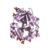
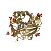
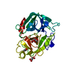
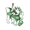
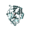
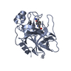
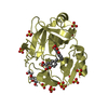
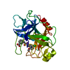
| Title | High resolution structure of Human Kallikrein 7 in Complex with Suc-Ala-Ala-Pro-Phe-chloromethylketone | ||||||
 Components Components | Kallikrein-7 | ||||||
 Keywords Keywords | HYDROLASE / S1 pocket / chloromethyl ketone / alternate conformations / Alternative splicing / Glycoprotein / Protease / Secreted / Serine protease / Zymogen | ||||||
| Function / homology |  Function and homology information Function and homology informationstratum corneum chymotryptic enzyme / positive regulation of antibacterial peptide production / epidermal lamellar body / cornified envelope / Differentiation of Keratinocytes in Interfollicular Epidermis in Mammalian Skin / extracellular matrix disassembly / epidermis development / Degradation of the extracellular matrix / serine-type peptidase activity / secretory granule ...stratum corneum chymotryptic enzyme / positive regulation of antibacterial peptide production / epidermal lamellar body / cornified envelope / Differentiation of Keratinocytes in Interfollicular Epidermis in Mammalian Skin / extracellular matrix disassembly / epidermis development / Degradation of the extracellular matrix / serine-type peptidase activity / secretory granule / protein maturation / metalloendopeptidase activity / peptidase activity / serine-type endopeptidase activity / proteolysis / extracellular space / extracellular region Similarity search - Function | ||||||
| Biological species |  Homo sapiens (human) Homo sapiens (human) | ||||||
| Method |  X-RAY DIFFRACTION / SYNCHROTRON / MOLECULAR REPLACEMENT / molecular replacement / Resolution: 1 Å X-RAY DIFFRACTION / SYNCHROTRON / MOLECULAR REPLACEMENT / molecular replacement / Resolution: 1 Å | ||||||
 Authors Authors | Debela, M. / Hess, P. / Magdolen, V. / Schechter, N.M. / Bode, W. / Goettig, P. | ||||||
 Citation Citation | Journal: Proc.Natl.Acad.Sci.Usa / Year: 2007 Title: Chymotryptic specificity determinants in the 1.0 A structure of the zinc-inhibited human tissue kallikrein 7. Authors: Debela, M. / Hess, P. / Magdolen, V. / Schechter, N.M. / Steiner, T. / Huber, R. / Bode, W. / Goettig, P. | ||||||
| History |
|
- Structure visualization
| Structure viewer | Molecule: MolmilJmol/JSmol |
|---|
- Downloads & links
-Download
| PDBx/mmCIF format | 2qxi.cif.gz | 117.3 KB | Display | PDBx/mmCIF format |
|---|---|---|---|---|
| PDB format | pdb2qxi.ent.gz | 88.7 KB | Display | PDB format |
| PDBx/mmJSON format | 2qxi.json.gz | Tree view | PDBx/mmJSON format | |
| Others |  Other downloads Other downloads |
-Validation report
| Summary document | 2qxi_validation.pdf.gz | 912.5 KB | Display | wwPDB validaton report |
|---|---|---|---|---|
| Full document | 2qxi_full_validation.pdf.gz | 914.7 KB | Display | |
| Data in XML | 2qxi_validation.xml.gz | 13.8 KB | Display | |
| Data in CIF | 2qxi_validation.cif.gz | 20.5 KB | Display | |
| Arichive directory | https://data.pdbj.org/pub/pdb/validation_reports/qx/2qxiftp://data.pdbj.org/pub/pdb/validation_reports/qx/2qxi | HTTPS FTP |
-Related structure data
| Related structure data |  2qxgC 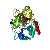 2qxhC 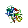 2qxjC 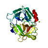 1lo6S C: citing same article ( S: Starting model for refinement |
|---|---|
| Similar structure data |


-Links
 PDBj
PDBj

- Assembly
| Deposited unit | 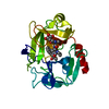
| ||||||||
|---|---|---|---|---|---|---|---|---|---|
| 1 |
| ||||||||
| Unit cell |
|
-Components
| #1: Protein | Mass: 24481.160 Da / Num. of mol.: 1 Source method: isolated from a genetically manipulated source Source: (gene. exp.) Homo sapiens (human) / Gene: KLK7, PRSS6, SCCE / Production host:   Spodoptera frugiperda (fall armyworm) / Strain (production host): SF9 Spodoptera frugiperda (fall armyworm) / Strain (production host): SF9References: UniProt: P49862, stratum corneum chymotryptic enzyme |
|---|---|
| #2: Chemical | ChemComp-K7J /   Mass: 539.021 Da / Num. of mol.: 1 / Source method: obtained synthetically / Formula: C25H35ClN4O7 Mass: 539.021 Da / Num. of mol.: 1 / Source method: obtained synthetically / Formula: C25H35ClN4O7 |
| #3: Water | ChemComp-HOH /  Mass: 18.015 Da / Num. of mol.: 225 / Source method: isolated from a natural source / Formula: H2O Mass: 18.015 Da / Num. of mol.: 225 / Source method: isolated from a natural source / Formula: H2O |
| Has protein modification | Y |
-Experimental details
-Experiment
| Experiment | Method: X-RAY DIFFRACTION / Number of used crystals: 1 |
|---|
- Sample preparation
| Crystal | Density Matthews: 1.99 Å3/Da / Density % sol: 38.08 % |
|---|---|
| Crystal grow | Temperature: 291 K / Method: vapor diffusion, sitting drop / pH: 6.5 Details: 30% MPD, 0.1 M sodium cacodylate, 0.2 M magnesium acetate, pH 6.5, VAPOR DIFFUSION, SITTING DROP, temperature 291K |
-Data collection
| Diffraction | Mean temperature: 100 K |
|---|---|
| Diffraction source | Source: SYNCHROTRON / Site: MPG/DESY, HAMBURG  / Beamline: BW6 / Wavelength: 1.05 Å / Beamline: BW6 / Wavelength: 1.05 Å |
| Detector | Type: MAR CCD 165 mm / Detector: CCD / Date: Jul 30, 2005 / Details: mirrors |
| Radiation | Monochromator: Si 111 CHANNEL / Protocol: SINGLE WAVELENGTH / Monochromatic (M) / Laue (L): M / Scattering type: x-ray |
| Radiation wavelength | Wavelength: 1.05 Å / Relative weight: 1 |
| Reflection | Resolution: 0.999→44.64 Å / Num. all: 97071 / Num. obs: 97071 / % possible obs: 94 % / Observed criterion σ(I): 0 / Redundancy: 3.3 % / Biso Wilson estimate: 12.3 Å2 / Rmerge(I) obs: 0.035 / Rsym value: 0.035 / Net I/σ(I): 9.7 |
| Reflection shell | Resolution: 1→1.05 Å / Redundancy: 2.9 % / Rmerge(I) obs: 0.28 / Mean I/σ(I) obs: 2.4 / Num. measured all: 38559 / Num. unique all: 13473 / Rsym value: 0.28 / % possible all: 89.6 |
-Phasing
| Phasing | Method: molecular replacement |
|---|
- Processing
| Software |
| |||||||||||||||||||||||||||||||||
|---|---|---|---|---|---|---|---|---|---|---|---|---|---|---|---|---|---|---|---|---|---|---|---|---|---|---|---|---|---|---|---|---|---|---|
| Refinement | Method to determine structure: MOLECULAR REPLACEMENT Starting model: 1LO6 Resolution: 1→10 Å / Num. parameters: 18790 / Num. restraintsaints: 23566 / Cross valid method: FREE R / σ(I): 0 / Stereochemistry target values: ENGH AND HUBER Details: ANISOTROPIC REFINEMENT REDUCED FREE R (NO CUTOFF), riding hydrogen model of SHELX was employed in the refinement
| |||||||||||||||||||||||||||||||||
| Solvent computation | Solvent model: MOEWS & KRETSINGER, J.MOL.BIOL.91(1973)201-228 | |||||||||||||||||||||||||||||||||
| Displacement parameters | Biso mean: 16.863 Å2 | |||||||||||||||||||||||||||||||||
| Refine analyze | Luzzati coordinate error obs: 0.089 Å | |||||||||||||||||||||||||||||||||
| Refinement step | Cycle: LAST / Resolution: 1→10 Å
| |||||||||||||||||||||||||||||||||
| Refine LS restraints |
| |||||||||||||||||||||||||||||||||
| LS refinement shell | Resolution: 1→1.1 Å /
|
