spontaneous exocytosis of neurotransmitter / negative regulation of meiotic cell cycle / PKA activation in glucagon signalling / CREB1 phosphorylation through the activation of Adenylate Cyclase / HDL assembly / regulation of protein processing / DARPP-32 events / Rap1 signalling / regulation of protein kinase activity / PKA activation ...spontaneous exocytosis of neurotransmitter / negative regulation of meiotic cell cycle / PKA activation in glucagon signalling / CREB1 phosphorylation through the activation of Adenylate Cyclase / HDL assembly / regulation of protein processing / DARPP-32 events / Rap1 signalling / regulation of protein kinase activity / PKA activation / Regulation of insulin secretion / Vasopressin regulates renal water homeostasis via Aquaporins / cellular response to parathyroid hormone stimulus / GPER1 signaling / protein localization to lipid droplet / Glucagon-like Peptide-1 (GLP1) regulates insulin secretion / Factors involved in megakaryocyte development and platelet production / cAMP-dependent protein kinase regulator activity / Hedgehog 'off' state / Loss of Nlp from mitotic centrosomes / Recruitment of mitotic centrosome proteins and complexes / Loss of proteins required for interphase microtubule organization from the centrosome / Anchoring of the basal body to the plasma membrane / MAPK6/MAPK4 signaling / regulation of cellular respiration / Interleukin-3, Interleukin-5 and GM-CSF signaling / Recruitment of NuMA to mitotic centrosomes / CD209 (DC-SIGN) signaling / AURKA Activation by TPX2 / RET signaling / High laminar flow shear stress activates signaling by PIEZO1 and PECAM1:CDH5:KDR in endothelial cells / GLI3 is processed to GLI3R by the proteasome / Regulation of PLK1 Activity at G2/M Transition / Mitochondrial protein degradation / cellular response to cold / VEGFA-VEGFR2 Pathway / nucleotide-activated protein kinase complex / interleukin-2-mediated signaling pathway / Ion homeostasis / mesoderm formation / cAMP-dependent protein kinase inhibitor activity / TORC1 signaling / potassium channel inhibitor activity / histone H1-4S35 kinase activity / cAMP-dependent protein kinase / positive regulation of triglyceride catabolic process / negative regulation of glycolytic process / beta-2 adrenergic receptor binding / cAMP-dependent protein kinase activity / regulation of bicellular tight junction assembly / cAMP-dependent protein kinase complex / neural tube closure / negative regulation of glycolytic process through fructose-6-phosphate / sperm capacitation / regulation of osteoblast differentiation / peptidyl-threonine phosphorylation / protein kinase A regulatory subunit binding / ciliary base / protein kinase A catalytic subunit binding / small molecule binding / intracellular potassium ion homeostasis / plasma membrane raft / axoneme / cAMP/PKA signal transduction / postsynaptic modulation of chemical synaptic transmission / cAMP binding / sperm flagellum / regulation of synaptic transmission, glutamatergic / negative regulation of cAMP/PKA signal transduction / positive regulation of gluconeogenesis / cellular response to nutrient levels / positive regulation of protein export from nucleus / negative regulation of TORC1 signaling / protein serine/threonine/tyrosine kinase activity / cellular response to glucagon stimulus / negative regulation of smoothened signaling pathway / positive regulation of phagocytosis / T-tubule / acrosomal vesicle / sperm midpiece / regulation of proteasomal protein catabolic process / negative regulation of protein localization to chromatin / lipid droplet / cellular response to glucose stimulus / neuromuscular junction / regulation of microtubule cytoskeleton organization / positive regulation of cholesterol biosynthetic process / peptidyl-serine phosphorylation / modulation of chemical synaptic transmission / small GTPase binding / mRNA processing / adenylate cyclase-inhibiting G protein-coupled receptor signaling pathway / manganese ion binding / protein autophosphorylation / cellular response to heat / presynapse / adenylate cyclase-activating G protein-coupled receptor signaling pathway / regulation of cell cycle / protein kinase activity / protein phosphorylation Similarity search - Function
cAMP-dependent protein kinase regulatory subunit / cAMP-dependent protein kinase regulatory subunit, dimerization-anchoring domain / Regulatory subunit of type II PKA R-subunit / RIIalpha, Regulatory subunit portion of type II PKA R-subunit / : / Cyclic nucleotide-binding domain signature 2. / cAMP-dependent protein kinase catalytic subunit / Cyclic nucleotide-binding domain signature 1. / Cyclic nucleotide-binding, conserved site / Cyclic nucleotide-monophosphate binding domain ...cAMP-dependent protein kinase regulatory subunit / cAMP-dependent protein kinase regulatory subunit, dimerization-anchoring domain / Regulatory subunit of type II PKA R-subunit / RIIalpha, Regulatory subunit portion of type II PKA R-subunit / : / Cyclic nucleotide-binding domain signature 2. / cAMP-dependent protein kinase catalytic subunit / Cyclic nucleotide-binding domain signature 1. / Cyclic nucleotide-binding, conserved site / Cyclic nucleotide-monophosphate binding domain / Cyclic nucleotide-binding domain / cAMP/cGMP binding motif profile. / Cyclic nucleotide-binding domain / Jelly Rolls / Cyclic nucleotide-binding domain superfamily / Extension to Ser/Thr-type protein kinases / AGC-kinase, C-terminal / AGC-kinase C-terminal domain profile. / RmlC-like jelly roll fold / Phosphorylase Kinase; domain 1 / Phosphorylase Kinase; domain 1 / Transferase(Phosphotransferase) domain 1 / Transferase(Phosphotransferase); domain 1 / Jelly Rolls / Serine/threonine-protein kinase, active site / Serine/Threonine protein kinases active-site signature. / Protein kinase domain / Serine/Threonine protein kinases, catalytic domain / Protein kinase, ATP binding site / Protein kinases ATP-binding region signature. / Protein kinase domain profile. / Protein kinase domain / Protein kinase-like domain superfamily / Sandwich / 2-Layer Sandwich / Orthogonal Bundle / Mainly Beta / Mainly Alpha / Alpha Beta Similarity search - Domain/homology
cAMP-dependent protein kinase catalytic subunit alpha / cAMP-dependent protein kinase type II-alpha regulatory subunit Similarity search - Component
In the structure databanks used in Yorodumi, some data are registered as the other names, "COVID-19 virus" and "2019-nCoV". Here are the details of the virus and the list of structure data.
Jan 31, 2019. EMDB accession codes are about to change! (news from PDBe EMDB page)
EMDB accession codes are about to change! (news from PDBe EMDB page)
The allocation of 4 digits for EMDB accession codes will soon come to an end. Whilst these codes will remain in use, new EMDB accession codes will include an additional digit and will expand incrementally as the available range of codes is exhausted. The current 4-digit format prefixed with “EMD-” (i.e. EMD-XXXX) will advance to a 5-digit format (i.e. EMD-XXXXX), and so on. It is currently estimated that the 4-digit codes will be depleted around Spring 2019, at which point the 5-digit format will come into force.
The EM Navigator/Yorodumi systems omit the EMD- prefix.
Related info.:Q: What is EMD? / ID/Accession-code notation in Yorodumi/EM Navigator
Yorodumi is a browser for structure data from EMDB, PDB, SASBDB, etc.
This page is also the successor to EM Navigator detail page, and also detail information page/front-end page for Omokage search.
The word "yorodu" (or yorozu) is an old Japanese word meaning "ten thousand". "mi" (miru) is to see.
Related info.:EMDB / PDB / SASBDB / Comparison of 3 databanks / Yorodumi Search / Aug 31, 2016. New EM Navigator & Yorodumi / Yorodumi Papers / Jmol/JSmol / Function and homology information / Changes in new EM Navigator and Yorodumi
 Movie
Movie Controller

 Yorodumi
 Open data
 Basic information
 Components
 Keywords
 Function and homology information
 Mus musculus (house mouse)
 X-RAY DIFFRACTION /
  Authors
 Citation
 Structure visualization
 Downloads & links
 Other downloads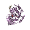
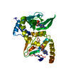

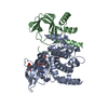
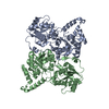

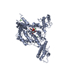
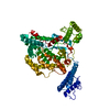
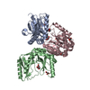
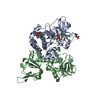

 PDBj
PDBj
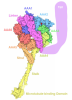
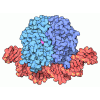


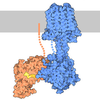


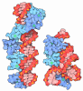
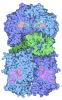
 Assembly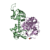

 Escherichia coli BL21(DE3) (bacteria) / Strain (production host): BL21(DE3) / References: UniProt: P05132, cAMP-dependent protein kinase
 Mass: 18.015 Da / Num. of mol.: 181 / Source method: isolated from a natural source / Formula: H2O
 Sample preparation
 / Beamline: 8.2.1 / Wavelength: 1 Å
 Processing