[English] 日本語
Yorodumi
- PDB-2qtq: Crystal structure of a predicted dna-binding transcriptional regu... -

+
Open data


ID or keywords:

Loading...

-
Basic information

Entry
Database: PDB / ID: 2qtq
TitleCrystal structure of a predicted dna-binding transcriptional regulator (saro_1072) from novosphingobium aromaticivorans dsm at 1.85 A resolution
ComponentsTranscriptional regulator, TetR family
KeywordsTRANSCRIPTION / Transcription regulator / dna/rna-binding 3-helical bundle fold / helix turn helix motif / hth motif / transcription regulation / structural genomics / Joint Center for Structural Genomics / JCSG / Protein Structure Initiative / PSI-2
Function / homology
Function and homology information


transcription cis-regulatory region binding / DNA-binding transcription factor activity
Similarity search - Function
Tetracyclin repressor-like, C-terminal domain / Tetracyclin repressor-like, C-terminal domain / : / Tetracycline Repressor, domain 2 / Tetracyclin repressor-like, C-terminal domain superfamily / Tetracycline Repressor; domain 2 / DNA-binding HTH domain, TetR-type / TetR-type HTH domain profile. / Homeobox-like domain superfamily / Orthogonal Bundle / Mainly Alpha
Similarity search - Domain/homology
NITRATE ION / Transcriptional regulator, TetR family
Similarity search - Component
Biological speciesNovosphingobium aromaticivorans (bacteria)
MethodX-RAY DIFFRACTION / SYNCHROTRON / MAD / Resolution: 1.85 Å
AuthorsJoint Center for Structural Genomics (JCSG)
CitationJournal: To be published
Title: Crystal structure of predicted DNA-binding transcriptional regulator (YP_496351.1) from Novosphingobium aromaticivorans DSM 12444 at 1.85 A resolution
Authors: Joint Center for Structural Genomics (JCSG)
History
DepositionAug 2, 2007Deposition site: RCSB / Processing site: RCSB
Revision 1.0Aug 21, 2007Provider: repository / Type: Initial release
Revision 1.1Jul 13, 2011Group: Advisory / Source and taxonomy / Version format compliance
Revision 1.2Oct 25, 2017Group: Author supporting evidence / Refinement description / Category: pdbx_struct_assembly_auth_evidence / software / Item: _software.classification / _software.name
Revision 1.3Jul 24, 2019Group: Data collection / Derived calculations / Refinement description
Category: software / struct_conn
Item: _software.classification / _software.contact_author ..._software.classification / _software.contact_author / _software.contact_author_email / _software.language / _software.location / _software.name / _software.type / _software.version / _struct_conn.pdbx_leaving_atom_flag
Revision 1.4Jan 25, 2023Group: Database references / Derived calculations / Category: database_2 / struct_ref_seq_dif / struct_site
Item: _database_2.pdbx_DOI / _database_2.pdbx_database_accession ..._database_2.pdbx_DOI / _database_2.pdbx_database_accession / _struct_ref_seq_dif.details / _struct_site.pdbx_auth_asym_id / _struct_site.pdbx_auth_comp_id / _struct_site.pdbx_auth_seq_id
Revision 1.5Oct 16, 2024Group: Data collection / Refinement description / Structure summary
Category: chem_comp_atom / chem_comp_bond ...chem_comp_atom / chem_comp_bond / pdbx_entry_details / pdbx_modification_feature / struct_ncs_dom_lim
Item: _struct_ncs_dom_lim.beg_auth_comp_id / _struct_ncs_dom_lim.end_auth_comp_id
Remark 300 
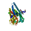
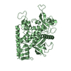
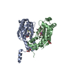
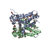
BIOMOLECULE: 1, 2 SEE REMARK 350 FOR THE AUTHOR PROVIDED AND PROGRAM GENERATED ASSEMBLY ... BIOMOLECULE: 1, 2 SEE REMARK 350 FOR THE AUTHOR PROVIDED AND PROGRAM GENERATED ASSEMBLY INFORMATION FOR THE STRUCTURE IN THIS ENTRY. THE REMARK MAY ALSO PROVIDE INFORMATION ON BURIED SURFACE AREA. SIZE EXCLUSION CHROMATOGRAPHY SUPPORTS THE ASSIGNMENT OF A DIMER AS A BIOLOGICALLY SIGNIFICANT OLIGOMERIZATION STATE.
Remark 999 SEQUENCE THE CONSTRUCT WAS EXPRESSED WITH A PURIFICATION TAG MGSDKIHHHHHHENLYFQG. THE TAG WAS ... SEQUENCE THE CONSTRUCT WAS EXPRESSED WITH A PURIFICATION TAG MGSDKIHHHHHHENLYFQG. THE TAG WAS REMOVED WITH TEV PROTEASE LEAVING ONLY A GLYCINE FOLLOWED BY THE TARGET SEQUENCE.

-
Structure visualization

Structure viewerMolecule:
MolmilJmol/JSmol

Downloads & links

-
Assembly

Deposited unit
A: Transcriptional regulator, TetR family
B: Transcriptional regulator, TetR family
C: Transcriptional regulator, TetR family
D: Transcriptional regulator, TetR family
hetero molecules


Theoretical massNumber of molelcules
Total (without water)99,61631
Polymers97,9404
Non-polymers1,67627
Water12,106672
1
A: Transcriptional regulator, TetR family
B: Transcriptional regulator, TetR family
hetero molecules


Theoretical massNumber of molelcules
Total (without water)50,14921
Polymers48,9702
Non-polymers1,17919
Water362
TypeNameSymmetry operationNumber
identity operation1_555x,y,z1
Buried area6190 Å2
MethodPISA
2
C: Transcriptional regulator, TetR family
D: Transcriptional regulator, TetR family
hetero molecules


Theoretical massNumber of molelcules
Total (without water)49,46710
Polymers48,9702
Non-polymers4968
Water362
TypeNameSymmetry operationNumber
identity operation1_555x,y,z1
Buried area5870 Å2
MethodPISA
Unit cell
Length a, b, c (Å)66.465, 98.762, 164.039
Angle α, β, γ (deg.)90.000, 90.000, 90.000
Int Tables number19
Space group name H-MP212121
Noncrystallographic symmetry (NCS)NCS domain:
IDEns-IDDetails
11A
21B
31C
41D

NCS domain segments:

Component-ID: 1 / Ens-ID: 1 / Beg auth comp-ID: GLY / Beg label comp-ID: GLY / End auth comp-ID: LEU / End label comp-ID: LEU / Refine code: 6 / Auth seq-ID: 15 - 210 / Label seq-ID: 16 - 211

Dom-IDAuth asym-IDLabel asym-ID
1AA
2BB
3CC
4DD
DetailsSIZE EXCLUSION CHROMATOGRAPHY SUPPORTS THE ASSIGNMENT OF A DIMER AS A BIOLOGICALLY SIGNIFICANT OLIGOMERIZATION STATE.

-
Components

#1: Protein
Transcriptional regulator, TetR family


Mass: 24485.025 Da / Num. of mol.: 4
Source method: isolated from a genetically manipulated source
Source: (gene. exp.) Novosphingobium aromaticivorans (bacteria)
Strain: DSM 12444 / Gene: YP_496351.1, Saro_1072 / Plasmid: speedET / Production host: Escherichia coli (E. coli) / Strain (production host): HK100 / References: UniProt: Q2G9F6
#2: Chemical ChemComp-NO3 / NITRATE ION


Mass: 62.005 Da / Num. of mol.: 3 / Source method: obtained synthetically / Formula: NO3
#3: Chemical...
ChemComp-EDO / 1,2-ETHANEDIOL / ETHYLENE GLYCOL


Mass: 62.068 Da / Num. of mol.: 24 / Source method: obtained synthetically / Formula: C2H6O2
#4: Water ChemComp-HOH / water


Mass: 18.015 Da / Num. of mol.: 672 / Source method: isolated from a natural source / Formula: H2O
Has protein modificationY

-
Experimental details

-
Experiment

ExperimentMethod: X-RAY DIFFRACTION / Number of used crystals: 1

-
Sample preparation

CrystalDensity Matthews: 2.75 Å3/Da / Density % sol: 55.25 %
Crystal growTemperature: 277 K / Method: vapor diffusion, sitting drop / pH: 6.9
Details: NANODROP, 0.2M K2NO3, 20.0% PEG 3350, No Buffer pH 6.9, VAPOR DIFFUSION, SITTING DROP, temperature 277K

-
Data collection

DiffractionMean temperature: 100 K
Diffraction sourceSource: SYNCHROTRON / Site: SSRL / Beamline: BL11-1 / Wavelength: 0.91837, 0.97926, 0.97870
DetectorType: MARMOSAIC 325 mm CCD / Detector: CCD / Date: Jun 30, 2007 / Details: Flat mirror (vertical focusing)
RadiationMonochromator: Single crystal Si(111) bent (horizontal focusing)
Protocol: MAD / Monochromatic (M) / Laue (L): M / Scattering type: x-ray
Radiation wavelength
IDWavelength (Å)Relative weight
10.918371
20.979261
30.97871
ReflectionResolution: 1.85→29.501 Å / Num. obs: 91044 / % possible obs: 98.3 % / Redundancy: 4.2 % / Biso Wilson estimate: 31.38 Å2 / Rmerge(I) obs: 0.059 / Rsym value: 0.059 / Net I/σ(I): 14.2
Reflection shell

Diffraction-ID: 1

Resolution (Å)Redundancy (%)Rmerge(I) obsMean I/σ(I) obsNum. measured allNum. unique allRsym value% possible all
1.85-1.94.20.7471.62810466190.74797.4
1.9-1.954.20.5121.52722364360.51297.5
1.95-2.014.20.4051.92653762600.40597.7
2.01-2.074.20.2942.62581161030.29497.9
2.07-2.144.20.2233.42513759330.22397.9
2.14-2.214.20.1724.42434857590.17298.1
2.21-2.294.20.14752350255760.14798.3
2.29-2.394.20.1166.22252453440.11698.3
2.39-2.494.20.172167051370.198.4
2.49-2.624.20.0838.32082849520.08398.6
2.62-2.764.20.079.81993547410.0798.8
2.76-2.934.20.06111874944750.0698.8
2.93-3.134.20.05311.81767242280.05399.1
3.13-3.384.20.04712.71649439620.04799.1
3.38-3.74.20.043141523836660.04399.3
3.7-4.144.10.03914.51362633120.03999.2
4.14-4.784.10.04214.21216229590.04299.4
4.78-5.8540.04213.9999325160.04299.4
5.85-8.273.90.04214.5770919970.04299.4
8.27-29.5013.60.03616.5382010690.03691.9

-
Phasing

PhasingMethod: MAD

-
Processing

Software
NameVersionClassificationNB
REFMAC5.2.0019refinement
PHENIXrefinement
SHARPphasing
MolProbity3beta29model building
SCALAdata scaling
PDB_EXTRACT3data extraction
MAR345CCDdata collection
MOSFLMdata reduction
RefinementMethod to determine structure: MAD / Resolution: 1.85→29.501 Å / Cor.coef. Fo:Fc: 0.965 / Cor.coef. Fo:Fc free: 0.953 / SU B: 6.431 / SU ML: 0.097 / TLS residual ADP flag: LIKELY RESIDUAL / Cross valid method: THROUGHOUT / σ(F): 0 / ESU R: 0.127 / ESU R Free: 0.12
Stereochemistry target values: MAXIMUM LIKELIHOOD WITH PHASES
Details: 1. HYDROGENS HAVE BEEN ADDED IN THE RIDING POSITIONS. 2. ATOM RECORD CONTAINS RESIDUAL B FACTORS ONLY. 3. A MET-INHIBITION PROTOCOL WAS USED FOR SELENOMETHIONINE INCORPORATION DURING PROTEIN ...Details: 1. HYDROGENS HAVE BEEN ADDED IN THE RIDING POSITIONS. 2. ATOM RECORD CONTAINS RESIDUAL B FACTORS ONLY. 3. A MET-INHIBITION PROTOCOL WAS USED FOR SELENOMETHIONINE INCORPORATION DURING PROTEIN EXPRESSION. THE OCCUPANCY OF THE SE ATOMS IN THE MSE RESIDUES WAS REDUCED TO 0.75 TO ACCOUNT FOR THE REDUCED SCATTERING POWER DUE TO PARTIAL S-MET INCORPORATION. 4. NO3 IONS FROM THE CRYSTALLIZATION BUFFER AND ETHYLENE GLYCOL (EDO) FROM THE CRYO SOLUTION WERE MODELED INTO THE STRUCTURE. 5. UNEXPLAINED ELECTRON DENSITY NEAR RESIDUE 184 IN THE A SUBUNIT WAS NOT MODELED.
RfactorNum. reflection% reflectionSelection details
Rfree0.216 4544 5 %RANDOM
Rwork0.185 ---
obs0.186 91002 97.98 %-
Solvent computationIon probe radii: 0.8 Å / Shrinkage radii: 0.8 Å / VDW probe radii: 1.2 Å / Solvent model: MASK
Displacement parametersBiso mean: 30.383 Å2
Baniso -1Baniso -2Baniso -3
1--1.96 Å20 Å20 Å2
2--2.95 Å20 Å2
3----0.99 Å2
Refinement stepCycle: LAST / Resolution: 1.85→29.501 Å
ProteinNucleic acidLigandSolventTotal
Num. atoms6313 0 108 672 7093
Refine LS restraints
Refine-IDTypeDev idealDev ideal targetNumber
X-RAY DIFFRACTIONr_bond_refined_d0.0150.0226846
X-RAY DIFFRACTIONr_bond_other_d0.0010.024817
X-RAY DIFFRACTIONr_angle_refined_deg1.2411.989230
X-RAY DIFFRACTIONr_angle_other_deg0.912311603
X-RAY DIFFRACTIONr_dihedral_angle_1_deg5.3175857
X-RAY DIFFRACTIONr_dihedral_angle_2_deg31.31922.355327
X-RAY DIFFRACTIONr_dihedral_angle_3_deg13.321151199
X-RAY DIFFRACTIONr_dihedral_angle_4_deg16.9641579
X-RAY DIFFRACTIONr_chiral_restr0.0750.21021
X-RAY DIFFRACTIONr_gen_planes_refined0.0050.027726
X-RAY DIFFRACTIONr_gen_planes_other0.0020.021524
X-RAY DIFFRACTIONr_nbd_refined0.220.21711
X-RAY DIFFRACTIONr_nbd_other0.1930.25312
X-RAY DIFFRACTIONr_nbtor_refined0.1790.23566
X-RAY DIFFRACTIONr_nbtor_other0.0850.23583
X-RAY DIFFRACTIONr_xyhbond_nbd_refined0.1550.2546
X-RAY DIFFRACTIONr_symmetry_vdw_refined0.2020.227
X-RAY DIFFRACTIONr_symmetry_vdw_other0.1740.269
X-RAY DIFFRACTIONr_symmetry_hbond_refined0.1440.232
X-RAY DIFFRACTIONr_mcbond_it1.96534565
X-RAY DIFFRACTIONr_mcbond_other0.47831676
X-RAY DIFFRACTIONr_mcangle_it2.51856705
X-RAY DIFFRACTIONr_scbond_it4.80582867
X-RAY DIFFRACTIONr_scangle_it6.062112525
Refine LS restraints NCS

Ens-ID: 1 / Number: 2130 / Refine-ID: X-RAY DIFFRACTION

Dom-IDAuth asym-IDTypeRms dev position (Å)Weight position
1ALOOSE POSITIONAL1.395
2BLOOSE POSITIONAL1.995
3CLOOSE POSITIONAL1.275
4DLOOSE POSITIONAL1.235
1ALOOSE THERMAL4.0710
2BLOOSE THERMAL3.910
3CLOOSE THERMAL3.4910
4DLOOSE THERMAL3.8510
LS refinement shellResolution: 1.85→1.898 Å / Total num. of bins used: 20
RfactorNum. reflection% reflection
Rfree0.365 312 -
Rwork0.291 6304 -
obs-6616 97.12 %
Refinement TLS params.

Method: refined / Refine-ID: X-RAY DIFFRACTION

IDL112)L122)L132)L222)L232)L332)S11 (Å °)S12 (Å °)S13 (Å °)S21 (Å °)S22 (Å °)S23 (Å °)S31 (Å °)S32 (Å °)S33 (Å °)T112)T122)T132)T222)T232)T332)Origin x (Å)Origin y (Å)Origin z (Å)
10.49190.0242-0.20681.1692-0.20690.88870.0042-0.0563-0.09210.1479-0.01440.1430.0349-0.05520.0101-0.0506-0.01080.0227-0.06410.0033-0.055263.157278.897930.8658
20.8688-0.0954-0.30890.38680.09081.13660.06590.03130.1546-0.0202-0.0017-0.0999-0.22920.0839-0.0642-0.0517-0.02130.026-0.1486-0.0155-0.057175.763292.883517.6244
33.19380.5692-0.4240.918-0.43012.3689-0.05020.18350.54730.0876-0.00530.0227-0.430.09250.0555-0.0484-0.0168-0.0443-0.09220.0146-0.03656.221996.1917-8.9463
41.7499-0.22480.5341.07930.30582.4641-0.10890.3680.197-0.09580.0352-0.1872-0.24350.50850.0737-0.1128-0.07130.01810.12360.1222-0.041369.350688.634-26.3124
Refinement TLS group
IDRefine-IDRefine TLS-IDAuth asym-IDLabel asym-IDAuth seq-IDLabel seq-ID
1X-RAY DIFFRACTION1AA9 - 21210 - 213
2X-RAY DIFFRACTION2BB15 - 21116 - 212
3X-RAY DIFFRACTION3CC15 - 21116 - 212
4X-RAY DIFFRACTION4DD15 - 21216 - 213

+
About Yorodumi

-
News

-
Feb 9, 2022. New format data for meta-information of EMDB entries

New format data for meta-information of EMDB entries

  • Version 3 of the EMDB header file is now the official format.
  • The previous official version 1.9 will be removed from the archive.

Related info.:EMDB header

External links:wwPDB to switch to version 3 of the EMDB data model

-
Aug 12, 2020. Covid-19 info

Covid-19 info

URL: https://pdbj.org/emnavi/covid19.php

New page: Covid-19 featured information page in EM Navigator.

Related info.:Covid-19 info / Mar 5, 2020. Novel coronavirus structure data

+
Mar 5, 2020. Novel coronavirus structure data

Novel coronavirus structure data

Related info.:Yorodumi Speices / Aug 12, 2020. Covid-19 info

External links:COVID-19 featured content - PDBj / Molecule of the Month (242):Coronavirus Proteases

+
Jan 31, 2019. EMDB accession codes are about to change! (news from PDBe EMDB page)

EMDB accession codes are about to change! (news from PDBe EMDB page)

  • The allocation of 4 digits for EMDB accession codes will soon come to an end. Whilst these codes will remain in use, new EMDB accession codes will include an additional digit and will expand incrementally as the available range of codes is exhausted. The current 4-digit format prefixed with “EMD-” (i.e. EMD-XXXX) will advance to a 5-digit format (i.e. EMD-XXXXX), and so on. It is currently estimated that the 4-digit codes will be depleted around Spring 2019, at which point the 5-digit format will come into force.
  • The EM Navigator/Yorodumi systems omit the EMD- prefix.

Related info.:Q: What is EMD? / ID/Accession-code notation in Yorodumi/EM Navigator

External links:EMDB Accession Codes are Changing Soon! / Contact to PDBj

+
Jul 12, 2017. Major update of PDB

Major update of PDB

  • wwPDB released updated PDB data conforming to the new PDBx/mmCIF dictionary.
  • This is a major update changing the version number from 4 to 5, and with Remediation, in which all the entries are updated.
  • In this update, many items about electron microscopy experimental information are reorganized (e.g. em_software).
  • Now, EM Navigator and Yorodumi are based on the updated data.

External links:wwPDB Remediation / Enriched Model Files Conforming to OneDep Data Standards Now Available in the PDB FTP Archive

-
Yorodumi

Thousand views of thousand structures

  • Yorodumi is a browser for structure data from EMDB, PDB, SASBDB, etc.
  • This page is also the successor to EM Navigator detail page, and also detail information page/front-end page for Omokage search.
  • The word "yorodu" (or yorozu) is an old Japanese word meaning "ten thousand". "mi" (miru) is to see.

Related info.:EMDB / PDB / SASBDB / Comparison of 3 databanks / Yorodumi Search / Aug 31, 2016. New EM Navigator & Yorodumi / Yorodumi Papers / Jmol/JSmol / Function and homology information / Changes in new EM Navigator and Yorodumi

Read more