 Movie
Movie Controller

[English] 日本語
 Yorodumi
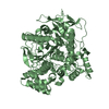
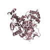
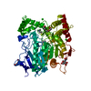
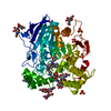
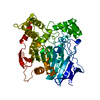
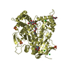
- PDB-1maa: MOUSE ACETYLCHOLINESTERASE CATALYTIC DOMAIN, GLYCOSYLATED PROTEIN -
+ Open data

- Basic information
| Entry | Database: PDB / ID: 1maa | |||||||||
|---|---|---|---|---|---|---|---|---|---|---|
| Title | MOUSE ACETYLCHOLINESTERASE CATALYTIC DOMAIN, GLYCOSYLATED PROTEIN | |||||||||
 Components Components | ACETYLCHOLINESTERASE | |||||||||
 Keywords Keywords | HYDROLASE / SERINE ESTERASE / ACETYLCHOLINESTERASE / TETRAMER / HYDROLASE FOLD / GLYCOSYLATED PROTEIN | |||||||||
| Function / homology |  Function and homology information Function and homology informationacetylcholine metabolic process / serine hydrolase activity / acetylcholine catabolic process / acetylcholinesterase / positive regulation of dendrite morphogenesis / cholinesterase activity / choline metabolic process / acetylcholine binding / osteoblast development / acetylcholine receptor signaling pathway ...acetylcholine metabolic process / serine hydrolase activity / acetylcholine catabolic process / acetylcholinesterase / positive regulation of dendrite morphogenesis / cholinesterase activity / choline metabolic process / acetylcholine binding / osteoblast development / acetylcholine receptor signaling pathway / acetylcholinesterase activity / positive regulation of axonogenesis / basement membrane / regulation of receptor recycling / side of membrane / synaptic cleft / collagen binding / laminin binding / synapse assembly / neuromuscular junction / response to insulin / receptor internalization / nuclear envelope / positive regulation of cold-induced thermogenesis / retina development in camera-type eye / presynaptic membrane / postsynaptic membrane / cell adhesion / membrane raft / endoplasmic reticulum lumen / axon / hydrolase activity / neuronal cell body / synapse / dendrite / perinuclear region of cytoplasm / cell surface / Golgi apparatus / protein homodimerization activity / : / extracellular region / membrane / identical protein binding / plasma membrane Similarity search - Function | |||||||||
| Biological species |  | |||||||||
| Method |  X-RAY DIFFRACTION / SYNCHROTRON / MOLECULAR REPLACEMENT / Resolution: 2.9 Å X-RAY DIFFRACTION / SYNCHROTRON / MOLECULAR REPLACEMENT / Resolution: 2.9 Å | |||||||||
 Authors Authors | Bourne, Y. / Taylor, P. / Bougis, P.E. / Marchot, P. | |||||||||
 Citation Citation | Journal: J.Biol.Chem. / Year: 1999 Title: Crystal structure of mouse acetylcholinesterase. A peripheral site-occluding loop in a tetrameric assembly. Authors: Bourne, Y. / Taylor, P. / Bougis, P.E. / Marchot, P. | |||||||||
| History |
|
- Structure visualization
| Structure viewer | Molecule: MolmilJmol/JSmol |
|---|
- Downloads & links
-Download
| PDBx/mmCIF format | 1maa.cif.gz | 419.3 KB | Display | PDBx/mmCIF format |
|---|---|---|---|---|
| PDB format | pdb1maa.ent.gz | 344.6 KB | Display | PDB format |
| PDBx/mmJSON format | 1maa.json.gz | Tree view | PDBx/mmJSON format | |
| Others |  Other downloads Other downloads |
-Validation report
| Arichive directory | https://data.pdbj.org/pub/pdb/validation_reports/ma/1maaftp://data.pdbj.org/pub/pdb/validation_reports/ma/1maa | HTTPS FTP |
|---|
-Related structure data
| Related structure data | 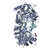 1mahS S: Starting model for refinement |
|---|---|
| Similar structure data |


-Links
 PDBj
PDBj
- Assembly
| Deposited unit | 
| ||||||||||||||||||||||||
|---|---|---|---|---|---|---|---|---|---|---|---|---|---|---|---|---|---|---|---|---|---|---|---|---|---|
| 1 | 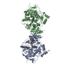
| ||||||||||||||||||||||||
| 2 | 
| ||||||||||||||||||||||||
| Unit cell |
| ||||||||||||||||||||||||
| Noncrystallographic symmetry (NCS) | NCS domain:
NCS oper:
|
-Components
-Protein , 1 types, 4 molecules ABCD
| #1: Protein | Mass: 60279.035 Da / Num. of mol.: 4 / Fragment: CATALYTIC DOMAIN Source method: isolated from a genetically manipulated source Source: (gene. exp.) Cell line (production host): HUMAN EMBRYONIC KIDNEY CELLS (HEK) Production host:  Homo sapiens (human) / References: UniProt: P21836, acetylcholinesterase Homo sapiens (human) / References: UniProt: P21836, acetylcholinesterase |
|---|
-Sugars , 2 types, 2 molecules 
| #2: Polysaccharide | 2-acetamido-2-deoxy-beta-D-glucopyranose-(1-4)-[beta-L-fucopyranose-(1-6)]2-acetamido-2-deoxy-beta- ...2-acetamido-2-deoxy-beta-D-glucopyranose-(1-4)-[beta-L-fucopyranose-(1-6)]2-acetamido-2-deoxy-beta-D-glucopyranose Source method: isolated from a genetically manipulated source |
|---|---|
| #3: Sugar | ChemComp-NAG /  Type: D-saccharide, beta linking / Mass: 221.208 Da / Num. of mol.: 1 Type: D-saccharide, beta linking / Mass: 221.208 Da / Num. of mol.: 1Source method: isolated from a genetically manipulated source Formula: C8H15NO6 |
-Non-polymers , 4 types, 198 molecules 


| #4: Chemical | ChemComp-DME /  Mass: 258.486 Da / Num. of mol.: 4 / Source method: obtained synthetically / Formula: C16H38N2 Mass: 258.486 Da / Num. of mol.: 4 / Source method: obtained synthetically / Formula: C16H38N2#5: Chemical | ChemComp-GOL /  Mass: 92.094 Da / Num. of mol.: 4 / Source method: obtained synthetically / Formula: C3H8O3 Mass: 92.094 Da / Num. of mol.: 4 / Source method: obtained synthetically / Formula: C3H8O3#6: Chemical |  Mass: 94.971 Da / Num. of mol.: 3 / Source method: obtained synthetically / Formula: PO4 Mass: 94.971 Da / Num. of mol.: 3 / Source method: obtained synthetically / Formula: PO4#7: Water | ChemComp-HOH / | Mass: 18.015 Da / Num. of mol.: 187 / Source method: isolated from a natural source / Formula: H2O |
|---|
-Details
| Has protein modification | Y |
|---|
-Experimental details
-Experiment
| Experiment | Method: X-RAY DIFFRACTION / Number of used crystals: 1 |
|---|
- Sample preparation
| Crystal | Density Matthews: 5.1 Å3/Da / Density % sol: 76 % | |||||||||||||||
|---|---|---|---|---|---|---|---|---|---|---|---|---|---|---|---|---|
| Crystal grow | pH: 7 / Details: pH 7.0 | |||||||||||||||
| Crystal grow | *PLUS Temperature: 4 ℃ / Method: vapor diffusion, hanging dropDetails: protein solution is mixed in a 1:1 ratio with well solution | |||||||||||||||
| Components of the solutions | *PLUS
|
-Data collection
| Diffraction | Mean temperature: 100 K |
|---|---|
| Diffraction source | Source: SYNCHROTRON / Site: EMBL/DESY, HAMBURG  / Beamline: X11 / Wavelength: 0.907 / Beamline: X11 / Wavelength: 0.907 |
| Detector | Type: MARRESEARCH / Detector: IMAGE PLATE / Date: Dec 1, 1996 / Details: BENT MIRROR |
| Radiation | Monochromator: GE(111) / Monochromatic (M) / Laue (L): M / Scattering type: x-ray |
| Radiation wavelength | Wavelength: 0.907 Å / Relative weight: 1 |
| Reflection | Resolution: 2.9→20 Å / Num. obs: 107379 / % possible obs: 92 % / Observed criterion σ(I): 0 / Redundancy: 2.8 % / Biso Wilson estimate: 55 Å2 / Rsym value: 0.105 / Net I/σ(I): 7 |
| Reflection shell | Resolution: 2.9→3 Å / Redundancy: 2.7 % / Mean I/σ(I) obs: 2 / Rsym value: 0.37 / % possible all: 83 |
| Reflection | *PLUS Num. measured all: 604052 / Rmerge(I) obs: 0.105 |
| Reflection shell | *PLUS % possible obs: 83 % / Rmerge(I) obs: 0.37 |
- Processing
| Software |
| ||||||||||||||||||||||||||||||||||||||||||||||||||||||||||||
|---|---|---|---|---|---|---|---|---|---|---|---|---|---|---|---|---|---|---|---|---|---|---|---|---|---|---|---|---|---|---|---|---|---|---|---|---|---|---|---|---|---|---|---|---|---|---|---|---|---|---|---|---|---|---|---|---|---|---|---|---|---|
| Refinement | Method to determine structure: MOLECULAR REPLACEMENT Starting model: 1MAH Resolution: 2.9→20 Å / Isotropic thermal model: RESTRAINED / Cross valid method: THROUGHOUT / σ(F): 0
| ||||||||||||||||||||||||||||||||||||||||||||||||||||||||||||
| Displacement parameters | Biso mean: 44 Å2 | ||||||||||||||||||||||||||||||||||||||||||||||||||||||||||||
| Refinement step | Cycle: LAST / Resolution: 2.9→20 Å
| ||||||||||||||||||||||||||||||||||||||||||||||||||||||||||||
| Refine LS restraints |
| ||||||||||||||||||||||||||||||||||||||||||||||||||||||||||||
| Refine LS restraints NCS | Refine-ID: X-RAY DIFFRACTION
| ||||||||||||||||||||||||||||||||||||||||||||||||||||||||||||
| LS refinement shell | Resolution: 2.9→2.97 Å / Total num. of bins used: 15
| ||||||||||||||||||||||||||||||||||||||||||||||||||||||||||||
| Xplor file |
| ||||||||||||||||||||||||||||||||||||||||||||||||||||||||||||
| Software | *PLUS Name: X-PLOR / Version: 3.8 / Classification: refinement | ||||||||||||||||||||||||||||||||||||||||||||||||||||||||||||
| Refinement | *PLUS | ||||||||||||||||||||||||||||||||||||||||||||||||||||||||||||
| Solvent computation | *PLUS | ||||||||||||||||||||||||||||||||||||||||||||||||||||||||||||
| Displacement parameters | *PLUS | ||||||||||||||||||||||||||||||||||||||||||||||||||||||||||||
| Refine LS restraints | *PLUS
| ||||||||||||||||||||||||||||||||||||||||||||||||||||||||||||
| LS refinement shell | *PLUS Rfactor obs: 0.32 |
