 Movie
Movie Controller


+ Open data

- Basic information
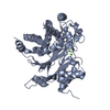
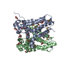
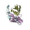
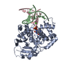
| Entry | Database: PDB / ID: 4m8b | ||||||
|---|---|---|---|---|---|---|---|
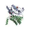
| Title | Fungal Protein | ||||||
 Components Components |
| ||||||
 Keywords Keywords | TRANSCRIPTION/DNA / WOPR fungal-pathogenesis transcription / WOPR domain / transcription factor / TRANSCRIPTION / TRANSCRIPTION-DNA complex | ||||||
| Function / homology | Gti1/Pac2 family / Gti1/Pac2 family / positive regulation of transcription by RNA polymerase II / DNA binding / nucleus / DNA / DNA (> 10) / Uncharacterized protein YHR177W Function and homology information Function and homology information | ||||||
| Biological species |  | ||||||
| Method |  X-RAY DIFFRACTION / SYNCHROTRON / MOLECULAR REPLACEMENT / Resolution: 2.61 Å X-RAY DIFFRACTION / SYNCHROTRON / MOLECULAR REPLACEMENT / Resolution: 2.61 Å | ||||||
 Authors Authors | Rosenberg, O.S. / Lohse, M.L. / Stroud, R.M. / Johnson, A.D. | ||||||
 Citation Citation | Journal: Proc.Natl.Acad.Sci.USA / Year: 2014 Title: Structure of a new DNA-binding domain which regulates pathogenesis in a wide variety of fungi. Authors: Lohse, M.B. / Rosenberg, O.S. / Cox, J.S. / Stroud, R.M. / Finer-Moore, J.S. / Johnson, A.D. | ||||||
| History |
|
- Structure visualization
| Structure viewer | Molecule: MolmilJmol/JSmol |
|---|
- Downloads & links
-Download
| PDBx/mmCIF format | 4m8b.cif.gz | 77.3 KB | Display | PDBx/mmCIF format |
|---|---|---|---|---|
| PDB format | pdb4m8b.ent.gz | 53.7 KB | Display | PDB format |
| PDBx/mmJSON format | 4m8b.json.gz | Tree view | PDBx/mmJSON format | |
| Others |  Other downloads Other downloads |
-Validation report
| Arichive directory | https://data.pdbj.org/pub/pdb/validation_reports/m8/4m8bftp://data.pdbj.org/pub/pdb/validation_reports/m8/4m8b | HTTPS FTP |
|---|
-Related structure data
| Similar structure data |
|---|


-Links
 PDBj
PDBj


- Assembly
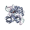
| Deposited unit | 
| ||||||||
|---|---|---|---|---|---|---|---|---|---|
| 1 |
| ||||||||
| Unit cell |
|
-Components
| #1: DNA chain | Mass: 6196.987 Da / Num. of mol.: 1 / Fragment: UNP residues 6-201 / Source method: obtained synthetically Details: DNA synthesis through standard methods and annealing of double stranded DNA. |
|---|---|
| #2: DNA chain | Mass: 6072.946 Da / Num. of mol.: 1 / Source method: obtained synthetically Details: DNA synthesis through standard methods and annealing of double stranded DNA. |
| #3: Protein | Mass: 23404.328 Da / Num. of mol.: 1 Source method: isolated from a genetically manipulated source Details: a pET21 based vector with an N-terminal 8-his tag followed by a 3C protease cleavage site Source: (gene. exp.) Strain: S288c / Gene: YHR177W, Yhr177wp / Plasmid: H3C / Production host:  |
| #4: Chemical | ChemComp-EDO /   Mass: 62.068 Da / Num. of mol.: 1 / Source method: obtained synthetically / Formula: C2H6O2 Mass: 62.068 Da / Num. of mol.: 1 / Source method: obtained synthetically / Formula: C2H6O2 |
| #5: Water | ChemComp-HOH /  Mass: 18.015 Da / Num. of mol.: 92 / Source method: isolated from a natural source / Formula: H2O Mass: 18.015 Da / Num. of mol.: 92 / Source method: isolated from a natural source / Formula: H2O |
-Experimental details
-Experiment
| Experiment | Method: X-RAY DIFFRACTION / Number of used crystals: 1 |
|---|
- Sample preparation
| Crystal | Density Matthews: 4.79 Å3/Da / Density % sol: 74.31 % |
|---|---|
| Crystal grow | Temperature: 298 K / Method: vapor diffusion, hanging drop / pH: 7.5 Details: 2 uL drops were a 1 to 1 mixture of protein with a well solution of 9 mM Calcium chloride, 10 mM Spermine, 50 mM Sodium cacodylate pH 7, 4.5% Isopropanol, 3.5% Ethylene glycol. ...Details: 2 uL drops were a 1 to 1 mixture of protein with a well solution of 9 mM Calcium chloride, 10 mM Spermine, 50 mM Sodium cacodylate pH 7, 4.5% Isopropanol, 3.5% Ethylene glycol. Cryoprotection involved a 4 to 6 mixture of 25% Glucose/Ethylene Glycol and well solution., VAPOR DIFFUSION, HANGING DROP, temperature 298K |
-Data collection
| Diffraction | Mean temperature: 80 K |
|---|---|
| Diffraction source | Source: SYNCHROTRON / Site: ALS  / Beamline: 8.3.1 / Wavelength: 1.115869 Å / Beamline: 8.3.1 / Wavelength: 1.115869 Å |
| Detector | Type: ADSC QUANTUM 315r / Detector: CCD / Date: Jan 10, 2013 |
| Radiation | Protocol: SINGLE WAVELENGTH / Monochromatic (M) / Laue (L): M / Scattering type: x-ray |
| Radiation wavelength | Wavelength: 1.115869 Å / Relative weight: 1 |
| Reflection | Resolution: 2.61→68.012 Å / Num. all: 19969 / Num. obs: 19969 / % possible obs: 93.6 % / Redundancy: 3.6 % / Rsym value: 0.057 / Net I/σ(I): 9.7 |
| Reflection shell | Resolution: 2.61→2.75 Å / Redundancy: 3.4 % / Rmerge(I) obs: 0.512 / Mean I/σ(I) obs: 1.5 / Num. unique all: 2684 / Rsym value: 0.512 / % possible all: 87.9 |
- Processing
| Software |
| ||||||||||||||||||
|---|---|---|---|---|---|---|---|---|---|---|---|---|---|---|---|---|---|---|---|
| Refinement | Method to determine structure: MOLECULAR REPLACEMENT Starting model: The starting model was a poor initial model built from MAD phases of a SeMet derivative. Resolution: 2.61→38.25 Å
| ||||||||||||||||||
| Refinement step | Cycle: LAST / Resolution: 2.61→38.25 Å
| ||||||||||||||||||
| Refine LS restraints |
| ||||||||||||||||||
| LS refinement shell | Resolution: 2.61→2.6753 Å
|
