 Movie
Movie Controller

[English] 日本語
 Yorodumi
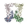
- PDB-2qs8: Crystal structure of a Xaa-Pro dipeptidase with bound methionine ... -
+ Open data

- Basic information
| Entry | Database: PDB / ID: 2qs8 | ||||||
|---|---|---|---|---|---|---|---|
| Title | Crystal structure of a Xaa-Pro dipeptidase with bound methionine in the active site | ||||||
 Components Components | Xaa-Pro Dipeptidase | ||||||
 Keywords Keywords | HYDROLASE / Amidohydrolase / Dipeptidase / TIM barrel / Protein Structure Initiative / PSI-2 / 9355e / NYSGXRC / Structural Genomics / New York SGX Research Center for Structural Genomics | ||||||
| Function / homology |  Function and homology information Function and homology informationhydrolase activity, acting on carbon-nitrogen (but not peptide) bonds / metal ion binding Similarity search - Function | ||||||
| Biological species |  Alteromonas macleodii (bacteria) Alteromonas macleodii (bacteria) | ||||||
| Method |  X-RAY DIFFRACTION / SYNCHROTRON / SAD / Resolution: 2.33 Å X-RAY DIFFRACTION / SYNCHROTRON / SAD / Resolution: 2.33 Å | ||||||
 Authors Authors | Kumaran, D. / Burley, S.K. / Swaminathan, S. / New York SGX Research Center for Structural Genomics (NYSGXRC) | ||||||
 Citation Citation | Journal: Biochemistry / Year: 2009 Title: Functional annotation of two new carboxypeptidases from the amidohydrolase superfamily of enzymes. Authors: Xiang, D.F. / Xu, C. / Kumaran, D. / Brown, A.C. / Sauder, J.M. / Burley, S.K. / Swaminathan, S. / Raushel, F.M. | ||||||
| History |
| ||||||
| Remark 999 | SEQUENCE THE SEQUENCE OF THIS PROTEIN WAS NOT AVAILABLE AT THE UNIPROT DATABASE AT THE TIME OF ... SEQUENCE THE SEQUENCE OF THIS PROTEIN WAS NOT AVAILABLE AT THE UNIPROT DATABASE AT THE TIME OF DEPOSITION. THE SEQUENCE INFORMATION IS AVAILABLE AT GENBANK WITH ACCESSION CODE GI:88795397. |
- Structure visualization
| Structure viewer | Molecule: MolmilJmol/JSmol |
|---|
- Downloads & links
-Download
| PDBx/mmCIF format | 2qs8.cif.gz | 168 KB | Display | PDBx/mmCIF format |
|---|---|---|---|---|
| PDB format | pdb2qs8.ent.gz | 134.5 KB | Display | PDB format |
| PDBx/mmJSON format | 2qs8.json.gz | Tree view | PDBx/mmJSON format | |
| Others |  Other downloads Other downloads |
-Validation report
| Arichive directory | https://data.pdbj.org/pub/pdb/validation_reports/qs/2qs8ftp://data.pdbj.org/pub/pdb/validation_reports/qs/2qs8 | HTTPS FTP |
|---|
-Related structure data
| Similar structure data | |
|---|---|
| Other databases |


-Links
 PDBj
PDBj

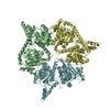
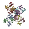
- Assembly
| Deposited unit | 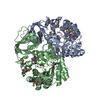
| ||||||||
|---|---|---|---|---|---|---|---|---|---|
| 1 | 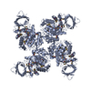
| ||||||||
| 2 | 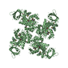
| ||||||||
| 3 | 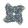
| ||||||||
| 4 |
| ||||||||
| Unit cell |
|
-Components
| #1: Protein | Mass: 46014.914 Da / Num. of mol.: 2 Source method: isolated from a genetically manipulated source Source: (gene. exp.) Alteromonas macleodii (bacteria) / Plasmid: pSGX3(BC) / Species (production host): Escherichia coli / Production host: #2: Chemical |   Mass: 24.305 Da / Num. of mol.: 2 / Source method: obtained synthetically / Formula: Mg Mass: 24.305 Da / Num. of mol.: 2 / Source method: obtained synthetically / Formula: Mg#3: Chemical |   Type: L-peptide linking / Mass: 149.211 Da / Num. of mol.: 2 / Source method: obtained synthetically / Formula: C5H11NO2S Type: L-peptide linking / Mass: 149.211 Da / Num. of mol.: 2 / Source method: obtained synthetically / Formula: C5H11NO2S#4: Water | ChemComp-HOH / |  Mass: 18.015 Da / Num. of mol.: 285 / Source method: isolated from a natural source / Formula: H2O Mass: 18.015 Da / Num. of mol.: 285 / Source method: isolated from a natural source / Formula: H2OHas protein modification | Y | |
|---|
-Experimental details
-Experiment
| Experiment | Method: X-RAY DIFFRACTION / Number of used crystals: 1 |
|---|
- Sample preparation
| Crystal | Density Matthews: 2.88 Å3/Da / Density % sol: 57.28 % |
|---|---|
| Crystal grow | Temperature: 298 K / pH: 7 Details: 30% MPD, 0.1M Hepes pH 7.0, 0.2M NaCl, VAPOR DIFFUSION, SITTING DROP, temperature 298K |
-Data collection
| Diffraction | Mean temperature: 100 K |
|---|---|
| Diffraction source | Source: SYNCHROTRON / Site: NSLS  / Beamline: X29A / Wavelength: 0.979 / Beamline: X29A / Wavelength: 0.979 |
| Detector | Type: ADSC QUANTUM 315 / Detector: CCD / Date: Jul 12, 2007 / Details: MIRRORS |
| Radiation | Monochromator: SI 111 / Protocol: SINGLE WAVELENGTH / Monochromatic (M) / Laue (L): M / Scattering type: x-ray |
| Radiation wavelength | Wavelength: 0.979 Å / Relative weight: 1 |
| Reflection | Resolution: 2.33→50 Å / Num. obs: 44488 / % possible obs: 99.8 % / Redundancy: 19 % / Biso Wilson estimate: 18.2 Å2 / Rmerge(I) obs: 0.1 / Net I/σ(I): 13 |
| Reflection shell | Resolution: 2.33→2.41 Å / Redundancy: 12 % / Rmerge(I) obs: 0.29 / Mean I/σ(I) obs: 3 / % possible all: 98.4 |
- Processing
| Software |
| ||||||||||||||||||||||||||||||||||||||||||||||||||||||||||||
|---|---|---|---|---|---|---|---|---|---|---|---|---|---|---|---|---|---|---|---|---|---|---|---|---|---|---|---|---|---|---|---|---|---|---|---|---|---|---|---|---|---|---|---|---|---|---|---|---|---|---|---|---|---|---|---|---|---|---|---|---|---|
| Refinement | Method to determine structure: SAD / Resolution: 2.33→45.81 Å / Rfactor Rfree error: 0.005 / Data cutoff high absF: 54537.76 / Data cutoff low absF: 0 / Isotropic thermal model: RESTRAINED / Cross valid method: THROUGHOUT / σ(F): 0 / Stereochemistry target values: ENGH & HUBER Details: RESIDUES LISTED AS MISSING IN REMARK 465 ARE DUE TO LACK OF ELECTRON DENSITY. RESIDUES WITH MISSING ATOMS LISTED IN REMARK 470 ARE DUE TO LACK OF ELECTRON DENSITY FOR SIDE CHAINS AND MODELED AS ALANINES.
| ||||||||||||||||||||||||||||||||||||||||||||||||||||||||||||
| Solvent computation | Solvent model: FLAT MODEL / Bsol: 21.08 Å2 / ksol: 0.31 e/Å3 | ||||||||||||||||||||||||||||||||||||||||||||||||||||||||||||
| Displacement parameters | Biso mean: 28.7 Å2
| ||||||||||||||||||||||||||||||||||||||||||||||||||||||||||||
| Refine analyze |
| ||||||||||||||||||||||||||||||||||||||||||||||||||||||||||||
| Refinement step | Cycle: LAST / Resolution: 2.33→45.81 Å
| ||||||||||||||||||||||||||||||||||||||||||||||||||||||||||||
| Refine LS restraints |
| ||||||||||||||||||||||||||||||||||||||||||||||||||||||||||||
| LS refinement shell | Resolution: 2.33→2.48 Å / Rfactor Rfree error: 0.014 / Total num. of bins used: 6
| ||||||||||||||||||||||||||||||||||||||||||||||||||||||||||||
| Xplor file |
|
