 Movie
Movie Controller

[English] 日本語
 Yorodumi
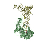
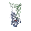
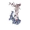
- PDB-2der: Cocrystal structure of an RNA sulfuration enzyme MnmA and tRNA-Gl... -
+ Open data

- Basic information
| Entry | Database: PDB / ID: 2der | ||||||
|---|---|---|---|---|---|---|---|
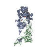
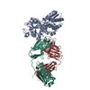
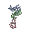
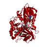
| Title | Cocrystal structure of an RNA sulfuration enzyme MnmA and tRNA-Glu in the initial tRNA binding state | ||||||
 Components Components |
| ||||||
 Keywords Keywords | Transferase/RNA / Protein-RNA complex / Transferase-RNA COMPLEX | ||||||
| Function / homology |  Function and homology information Function and homology informationtRNA-uridine 2-sulfurtransferase / tRNA-uridine 2-sulfurtransferase activity / stress response to acid chemical / sulfurtransferase activity / tRNA wobble position uridine thiolation / tRNA binding / ATP binding / cytosol Similarity search - Function | ||||||
| Biological species |  | ||||||
| Method |  X-RAY DIFFRACTION / SYNCHROTRON / MOLECULAR REPLACEMENT / Resolution: 3.1 Å X-RAY DIFFRACTION / SYNCHROTRON / MOLECULAR REPLACEMENT / Resolution: 3.1 Å | ||||||
 Authors Authors | Numata, T. / Ikeuchi, Y. / Fukai, S. / Suzuki, T. / Nureki, O. | ||||||
 Citation Citation | Journal: Nature / Year: 2006 Title: Snapshots of tRNA sulphuration via an adenylated intermediate Authors: Numata, T. / Ikeuchi, Y. / Fukai, S. / Suzuki, T. / Nureki, O. | ||||||
| History |
|
- Structure visualization
| Structure viewer | Molecule: MolmilJmol/JSmol |
|---|
- Downloads & links
-Download
| PDBx/mmCIF format | 2der.cif.gz | 230.6 KB | Display | PDBx/mmCIF format |
|---|---|---|---|---|
| PDB format | pdb2der.ent.gz | 179.7 KB | Display | PDB format |
| PDBx/mmJSON format | 2der.json.gz | Tree view | PDBx/mmJSON format | |
| Others |  Other downloads Other downloads |
-Validation report
| Arichive directory | https://data.pdbj.org/pub/pdb/validation_reports/de/2derftp://data.pdbj.org/pub/pdb/validation_reports/de/2der | HTTPS FTP |
|---|
-Related structure data
| Related structure data | 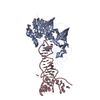 2detSC  2deuC S: Starting model for refinement C: citing same article ( |
|---|---|
| Similar structure data |


-Links
 PDBj
PDBj


- Assembly
| Deposited unit | 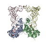
| ||||||||
|---|---|---|---|---|---|---|---|---|---|
| 1 | 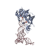
| ||||||||
| 2 | 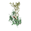
| ||||||||
| Unit cell |
|
-Components
| #1: RNA chain | Mass: 24378.514 Da / Num. of mol.: 2 / Source method: obtained synthetically / Details: RNA was prepared by in vitro transcription. #2: Protein | Mass: 42411.816 Da / Num. of mol.: 2 Source method: isolated from a genetically manipulated source Source: (gene. exp.) References: UniProt: P25745, Transferases; Transferring sulfur-containing groups; Sulfurtransferases #3: Chemical | ChemComp-PO4 /   Mass: 94.971 Da / Num. of mol.: 4 / Source method: obtained synthetically / Formula: PO4 Mass: 94.971 Da / Num. of mol.: 4 / Source method: obtained synthetically / Formula: PO4#4: Chemical | ChemComp-SO4 /   Mass: 96.063 Da / Num. of mol.: 5 / Source method: obtained synthetically / Formula: SO4 Mass: 96.063 Da / Num. of mol.: 5 / Source method: obtained synthetically / Formula: SO4Has protein modification | Y | |
|---|
-Experimental details
-Experiment
| Experiment | Method: X-RAY DIFFRACTION / Number of used crystals: 1 |
|---|
- Sample preparation
| Crystal | Density Matthews: 3.85 Å3/Da / Density % sol: 68.03 % Description: THE STRUCTURE FACTOR FILE CONTAINS FRIEDEL PAIRS. | ||||||||||||||||||||||||||||||||||||||||||||
|---|---|---|---|---|---|---|---|---|---|---|---|---|---|---|---|---|---|---|---|---|---|---|---|---|---|---|---|---|---|---|---|---|---|---|---|---|---|---|---|---|---|---|---|---|---|
| Crystal grow | Temperature: 293 K / Method: vapor diffusion, hanging drop / pH: 7.5 Details: 90mM HEPES-Na buffer (pH 7.5), 40mM ammonium dihydrogen phosphate, 1.8% PEG400, 1.7-1.9M ammonium sulfate, VAPOR DIFFUSION, HANGING DROP, temperature 293K | ||||||||||||||||||||||||||||||||||||||||||||
| Components of the solutions |
|
-Data collection
| Diffraction | Mean temperature: 30 K |
|---|---|
| Diffraction source | Source: SYNCHROTRON / Site: SPring-8  / Beamline: BL41XU / Wavelength: 0.97927 Å / Beamline: BL41XU / Wavelength: 0.97927 Å |
| Detector | Type: ADSC QUANTUM 315 / Detector: CCD / Date: Dec 7, 2004 |
| Radiation | Monochromator: Si 111 CHANNEL / Protocol: SINGLE WAVELENGTH / Monochromatic (M) / Laue (L): M / Scattering type: x-ray |
| Radiation wavelength | Wavelength: 0.97927 Å / Relative weight: 1 |
| Reflection | Resolution: 3.1→50 Å / Num. obs: 61680 / % possible obs: 89.8 % / Redundancy: 4.3 % / Biso Wilson estimate: 0 Å2 / Rmerge(I) obs: 0.118 / Net I/σ(I): 11.2 |
| Reflection shell | Resolution: 3.1→3.21 Å / Redundancy: 1.7 % / Rmerge(I) obs: 0.363 / Mean I/σ(I) obs: 1.8 / % possible all: 41.9 |
- Processing
| Software |
| ||||||||||||||||||||||||||||||||||||||||||||||||||||||||||||
|---|---|---|---|---|---|---|---|---|---|---|---|---|---|---|---|---|---|---|---|---|---|---|---|---|---|---|---|---|---|---|---|---|---|---|---|---|---|---|---|---|---|---|---|---|---|---|---|---|---|---|---|---|---|---|---|---|---|---|---|---|---|
| Refinement | Method to determine structure: MOLECULAR REPLACEMENT Starting model: PDB ENTRY 2DET Resolution: 3.1→46.75 Å / Rfactor Rfree error: 0.005 / Data cutoff high absF: 75333.9 / Data cutoff low absF: 0 / Isotropic thermal model: RESTRAINED / Cross valid method: THROUGHOUT / σ(F): 0 / Stereochemistry target values: Engh & Huber / Details: THE STRUCTURE FACTOR FILE CONTAINS FRIEDEL PAIRS.
| ||||||||||||||||||||||||||||||||||||||||||||||||||||||||||||
| Solvent computation | Solvent model: FLAT MODEL / Bsol: 24.9515 Å2 / ksol: 0.283117 e/Å3 | ||||||||||||||||||||||||||||||||||||||||||||||||||||||||||||
| Displacement parameters | Biso mean: 69 Å2
| ||||||||||||||||||||||||||||||||||||||||||||||||||||||||||||
| Refine analyze |
| ||||||||||||||||||||||||||||||||||||||||||||||||||||||||||||
| Refinement step | Cycle: LAST / Resolution: 3.1→46.75 Å
| ||||||||||||||||||||||||||||||||||||||||||||||||||||||||||||
| Refine LS restraints |
| ||||||||||||||||||||||||||||||||||||||||||||||||||||||||||||
| LS refinement shell | Resolution: 3.1→3.29 Å / Rfactor Rfree error: 0.031 / Total num. of bins used: 6
| ||||||||||||||||||||||||||||||||||||||||||||||||||||||||||||
| Xplor file |
|
