 Movie
Movie Controller


+ Open data

- Basic information
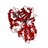
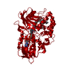
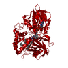
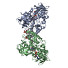
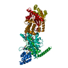
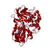
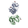
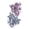
| Entry | Database: PDB / ID: 3nn0 | ||||||
|---|---|---|---|---|---|---|---|
| Title | Complex of 6-hydroxy-L-nicotine oxidase with nicotinamide | ||||||
 Components Components | 6-hydroxy-L-nicotine oxidase | ||||||
 Keywords Keywords | OXIDOREDUCTASE / ENANTHIOMERIC SUBSTRATE-INHIBITOR / FLAVOENZYMES / NICOTINE DEGRADATION / FAD fold / amino oxidase / FAD binding / cytosol | ||||||
| Function / homology |  Function and homology information Function and homology information(S)-6-hydroxynicotine oxidase / (S)-6-hydroxynicotine oxidase activity / nicotine catabolic process / alkaloid metabolic process / nucleotide binding / cytoplasm Similarity search - Function | ||||||
| Biological species |  Arthrobacter nicotinovorans (bacteria) Arthrobacter nicotinovorans (bacteria) | ||||||
| Method |  X-RAY DIFFRACTION / SYNCHROTRON / MOLECULAR REPLACEMENT / Resolution: 2.75 Å X-RAY DIFFRACTION / SYNCHROTRON / MOLECULAR REPLACEMENT / Resolution: 2.75 Å | ||||||
 Authors Authors | Kachalova, G.S. / Bartunik, H.D. | ||||||
 Citation Citation | Journal: J.Mol.Biol. / Year: 2010 Title: Crystal structure analysis of free and substrate-bound 6-hydroxy-L-nicotine oxidase from Arthrobacter nicotinovorans. Authors: Kachalova, G.S. / Bourenkov, G.P. / Mengesdorf, T. / Schenk, S. / Maun, H.R. / Burghammer, M. / Riekel, C. / Decker, K. / Bartunik, H.D. #1: Journal: J.Mol.Biol. / Year: 2010Title: Crystal structure analysis of free and substrate-bound 6-hydroxy-L-nicotine from Arthrobacter nicotinovorans Authors: Kachalova, G.S. / Bourenkov, G.P. / Mengesdorf, T. / Schenk, S. / Maun, H.R. / Burghammer, M. / Riekel, C. / Decker, K. / Bartunik, H.D. | ||||||
| History |
|
- Structure visualization
| Structure viewer | Molecule: MolmilJmol/JSmol |
|---|
- Downloads & links
-Download
| PDBx/mmCIF format | 3nn0.cif.gz | 106.1 KB | Display | PDBx/mmCIF format |
|---|---|---|---|---|
| PDB format | pdb3nn0.ent.gz | 79.2 KB | Display | PDB format |
| PDBx/mmJSON format | 3nn0.json.gz | Tree view | PDBx/mmJSON format | |
| Others |  Other downloads Other downloads |
-Validation report
| Arichive directory | https://data.pdbj.org/pub/pdb/validation_reports/nn/3nn0ftp://data.pdbj.org/pub/pdb/validation_reports/nn/3nn0 | HTTPS FTP |
|---|
-Related structure data
| Related structure data |  3k7mSC 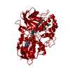 3k7qC  3k7tC 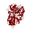 3ngcC 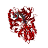 3nh3C 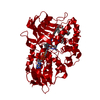 3nhoC  3nk0C  3nk1C 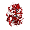 3nk2C  3nn6C C: citing same article ( S: Starting model for refinement |
|---|---|
| Similar structure data |


-Links
 PDBj
PDBj
- Assembly
| Deposited unit | 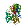
| ||||||||
|---|---|---|---|---|---|---|---|---|---|
| 1 | 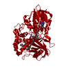
| ||||||||
| Unit cell |
|
-Components
| #1: Protein | Mass: 47214.090 Da / Num. of mol.: 1 Source method: isolated from a genetically manipulated source Source: (gene. exp.) Arthrobacter nicotinovorans (bacteria) / Gene: 6-HLNO / Plasmid: pTrc99A / Production host: |
|---|---|
| #2: Chemical | ChemComp-NCA / 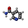  Mass: 122.125 Da / Num. of mol.: 1 / Source method: obtained synthetically / Formula: C6H6N2O / Comment: medication*YM Mass: 122.125 Da / Num. of mol.: 1 / Source method: obtained synthetically / Formula: C6H6N2O / Comment: medication*YM |
| #3: Chemical | ChemComp-FAD /   Mass: 785.550 Da / Num. of mol.: 1 / Source method: obtained synthetically / Formula: C27H33N9O15P2 / Comment: FAD*YM Mass: 785.550 Da / Num. of mol.: 1 / Source method: obtained synthetically / Formula: C27H33N9O15P2 / Comment: FAD*YM |
| #4: Chemical | ChemComp-GP7 / (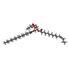  Mass: 673.901 Da / Num. of mol.: 1 / Source method: obtained synthetically / Formula: C36H68NO8P Mass: 673.901 Da / Num. of mol.: 1 / Source method: obtained synthetically / Formula: C36H68NO8P |
| #5: Water | ChemComp-HOH /  Mass: 18.015 Da / Num. of mol.: 234 / Source method: isolated from a natural source / Formula: H2O Mass: 18.015 Da / Num. of mol.: 234 / Source method: isolated from a natural source / Formula: H2O |
-Experimental details
-Experiment
| Experiment | Method: X-RAY DIFFRACTION / Number of used crystals: 1 |
|---|
- Sample preparation
| Crystal | Density Matthews: 3.95 Å3/Da / Density % sol: 68.87 % |
|---|---|
| Crystal grow | Temperature: 293 K / Method: vapor diffusion, sitting drop / pH: 7.5 Details: 20mM Sodium phosphate; 4M sodium formiate, pH 7.5, VAPOR DIFFUSION, SITTING DROP, temperature 293K |
-Data collection
| Diffraction | Mean temperature: 100 K |
|---|---|
| Diffraction source | Source: SYNCHROTRON / Site: MPG/DESY, HAMBURG  / Beamline: BW6 / Wavelength: 1.05 Å / Beamline: BW6 / Wavelength: 1.05 Å |
| Detector | Type: MAR CCD 165 mm / Detector: CCD / Date: Sep 8, 2009 / Details: mirror |
| Radiation | Monochromator: Graphite / Protocol: SINGLE WAVELENGTH / Monochromatic (M) / Laue (L): M / Scattering type: x-ray |
| Radiation wavelength | Wavelength: 1.05 Å / Relative weight: 1 |
| Reflection | Resolution: 2.75→20 Å / Num. all: 20443 / Num. obs: 20443 / % possible obs: 100 % / Observed criterion σ(I): 2 / Redundancy: 20 % / Biso Wilson estimate: 65 Å2 / Rmerge(I) obs: 0.157 / Net I/σ(I): 27.4 |
| Reflection shell | Resolution: 2.75→2.8 Å / Redundancy: 20 % / Rmerge(I) obs: 0.753 / Mean I/σ(I) obs: 3.7 / Num. unique all: 991 / % possible all: 100 |
- Processing
| Software |
| ||||||||||||||||||||||||||||||||||||||||||||||||||||||||||||||||||||||||||||||||||||||||||||||||||||||||||||||||||||||||||||||||||||||||||||||||||||||||||||||||||||||||||
|---|---|---|---|---|---|---|---|---|---|---|---|---|---|---|---|---|---|---|---|---|---|---|---|---|---|---|---|---|---|---|---|---|---|---|---|---|---|---|---|---|---|---|---|---|---|---|---|---|---|---|---|---|---|---|---|---|---|---|---|---|---|---|---|---|---|---|---|---|---|---|---|---|---|---|---|---|---|---|---|---|---|---|---|---|---|---|---|---|---|---|---|---|---|---|---|---|---|---|---|---|---|---|---|---|---|---|---|---|---|---|---|---|---|---|---|---|---|---|---|---|---|---|---|---|---|---|---|---|---|---|---|---|---|---|---|---|---|---|---|---|---|---|---|---|---|---|---|---|---|---|---|---|---|---|---|---|---|---|---|---|---|---|---|---|---|---|---|---|---|---|---|
| Refinement | Method to determine structure: MOLECULAR REPLACEMENT Starting model: PDB 3K7M Resolution: 2.75→14.4 Å / Cor.coef. Fo:Fc: 0.962 / Cor.coef. Fo:Fc free: 0.928 / SU B: 9.749 / SU ML: 0.195 / Isotropic thermal model: Isotropic / Cross valid method: THROUGHOUT / ESU R Free: 0.286 / Stereochemistry target values: MAXIMUM LIKELIHOOD
| ||||||||||||||||||||||||||||||||||||||||||||||||||||||||||||||||||||||||||||||||||||||||||||||||||||||||||||||||||||||||||||||||||||||||||||||||||||||||||||||||||||||||||
| Solvent computation | Ion probe radii: 0.8 Å / Shrinkage radii: 0.8 Å / VDW probe radii: 1.4 Å / Solvent model: BABINET MODEL WITH MASK | ||||||||||||||||||||||||||||||||||||||||||||||||||||||||||||||||||||||||||||||||||||||||||||||||||||||||||||||||||||||||||||||||||||||||||||||||||||||||||||||||||||||||||
| Displacement parameters | Biso mean: 44.823 Å2 | ||||||||||||||||||||||||||||||||||||||||||||||||||||||||||||||||||||||||||||||||||||||||||||||||||||||||||||||||||||||||||||||||||||||||||||||||||||||||||||||||||||||||||
| Refinement step | Cycle: LAST / Resolution: 2.75→14.4 Å
| ||||||||||||||||||||||||||||||||||||||||||||||||||||||||||||||||||||||||||||||||||||||||||||||||||||||||||||||||||||||||||||||||||||||||||||||||||||||||||||||||||||||||||
| Refine LS restraints |
| ||||||||||||||||||||||||||||||||||||||||||||||||||||||||||||||||||||||||||||||||||||||||||||||||||||||||||||||||||||||||||||||||||||||||||||||||||||||||||||||||||||||||||
| LS refinement shell | Resolution: 2.75→2.82 Å / Total num. of bins used: 20
|
