 Movie
Movie Controller

[English] 日本語
 Yorodumi
- PDB-2qnf: Crystal structure of T4 Endonuclease VII H43N mutant in complex w... -
+ Open data

- Basic information
| Entry | Database: PDB / ID: 2qnf | ||||||
|---|---|---|---|---|---|---|---|
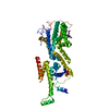
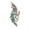
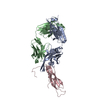
| Title | Crystal structure of T4 Endonuclease VII H43N mutant in complex with heteroduplex DNA containing base mismatches | ||||||
 Components Components |
| ||||||
 Keywords Keywords | Hydrolase/DNA / T4 Endonuclease VII / Endo VII / resolvase / resolving-enzyme / DNA mismatch / Alternative initiation / Calcium / Hydrolase / Metal-binding / Zinc / Hydrolase-DNA COMPLEX | ||||||
| Function / homology |  Function and homology information Function and homology informationendonuclease activity / Hydrolases; Acting on ester bonds / hydrolase activity / metal ion binding Similarity search - Function | ||||||
| Biological species |  Enterobacteria phage T4 (virus) Enterobacteria phage T4 (virus) | ||||||
| Method |  X-RAY DIFFRACTION / SYNCHROTRON / MOLECULAR REPLACEMENT / Resolution: 3 Å X-RAY DIFFRACTION / SYNCHROTRON / MOLECULAR REPLACEMENT / Resolution: 3 Å | ||||||
 Authors Authors | Biertumpfel, C. / Yang, W. / Suck, D. | ||||||
 Citation Citation | Journal: Nature / Year: 2007 Title: Crystal structure of T4 endonuclease VII resolving a Holliday junction. Authors: Biertumpfel, C. / Yang, W. / Suck, D. | ||||||
| History |
|
- Structure visualization
| Structure viewer | Molecule: MolmilJmol/JSmol |
|---|
- Downloads & links
-Download
| PDBx/mmCIF format | 2qnf.cif.gz | 111.3 KB | Display | PDBx/mmCIF format |
|---|---|---|---|---|
| PDB format | pdb2qnf.ent.gz | 81.6 KB | Display | PDB format |
| PDBx/mmJSON format | 2qnf.json.gz | Tree view | PDBx/mmJSON format | |
| Others |  Other downloads Other downloads |
-Validation report
| Arichive directory | https://data.pdbj.org/pub/pdb/validation_reports/qn/2qnfftp://data.pdbj.org/pub/pdb/validation_reports/qn/2qnf | HTTPS FTP |
|---|
-Related structure data
| Related structure data |  2qncC  1e7lS S: Starting model for refinement C: citing same article ( |
|---|---|
| Similar structure data |


-Links
 PDBj
PDBj


- Assembly
| Deposited unit | 
| ||||||||
|---|---|---|---|---|---|---|---|---|---|
| 1 |
| ||||||||
| Unit cell |
|
-Components
-DNA chain , 4 types, 4 molecules CDEF
| #1: DNA chain | Mass: 4908.193 Da / Num. of mol.: 1 / Source method: obtained synthetically |
|---|---|
| #2: DNA chain | Mass: 4890.166 Da / Num. of mol.: 1 / Source method: obtained synthetically |
| #3: DNA chain | Mass: 4890.166 Da / Num. of mol.: 1 / Source method: obtained synthetically |
| #4: DNA chain | Mass: 4908.193 Da / Num. of mol.: 1 / Source method: obtained synthetically |
-Protein / Non-polymers , 2 types, 4 molecules AB
| #5: Protein | Mass: 18149.732 Da / Num. of mol.: 2 / Mutation: H43N Source method: isolated from a genetically manipulated source Source: (gene. exp.) Enterobacteria phage T4 (virus) / Genus: T4-like viruses / Species: Enterobacteria phage T4 sensu lato / Gene: 49 / Plasmid: pET24d / Production host:  References: UniProt: P13340, crossover junction endodeoxyribonuclease #6: Chemical |  Mass: 65.409 Da / Num. of mol.: 2 / Source method: obtained synthetically / Formula: Zn Mass: 65.409 Da / Num. of mol.: 2 / Source method: obtained synthetically / Formula: Zn |
|---|
-Experimental details
-Experiment
| Experiment | Method: X-RAY DIFFRACTION / Number of used crystals: 1 |
|---|
- Sample preparation
| Crystal | Density Matthews: 3.35 Å3/Da / Density % sol: 63.23 % | ||||||||||||||||||||||||||||||||||||
|---|---|---|---|---|---|---|---|---|---|---|---|---|---|---|---|---|---|---|---|---|---|---|---|---|---|---|---|---|---|---|---|---|---|---|---|---|---|
| Crystal grow | Temperature: 293 K / pH: 6.5 Details: 25%(w/v) PEG 3350, 100 mM Bis-Tris, 220mM Li2SO4 and 5% (w/v) glucose, pH 6.5, VAPOR DIFFUSION, temperature 293K, pH 6.50 | ||||||||||||||||||||||||||||||||||||
| Components of the solutions |
|
-Data collection
| Diffraction | Mean temperature: 100 K |
|---|---|
| Diffraction source | Source: SYNCHROTRON / Site: APS  / Beamline: 22-ID / Wavelength: 1 / Beamline: 22-ID / Wavelength: 1 |
| Detector | Type: MARMOSAIC 300 mm CCD / Detector: CCD / Date: Aug 10, 2006 |
| Radiation | Monochromator: SI 220. ROSENBAUM-ROCK DOUBLE -CRYSTAL MONOCHROMATOR Protocol: SINGLE WAVELENGTH / Monochromatic (M) / Laue (L): M / Scattering type: x-ray |
| Radiation wavelength | Wavelength: 1 Å / Relative weight: 1 |
| Reflection | Resolution: 3→38 Å / Num. obs: 12214 / % possible obs: 84.6 % / Redundancy: 2.89 % / Biso Wilson estimate: 88.4 Å2 / Rsym value: 0.048 / Net I/σ(I): 20.8 |
| Reflection shell | Resolution: 3→3.11 Å / Mean I/σ(I) obs: 5.7 / Rsym value: 0.146 / % possible all: 37.6 |
- Processing
| Software |
| ||||||||||||||||||||||||||||||||||||||||||||||||||||||||||||||||||||||||||||||||
|---|---|---|---|---|---|---|---|---|---|---|---|---|---|---|---|---|---|---|---|---|---|---|---|---|---|---|---|---|---|---|---|---|---|---|---|---|---|---|---|---|---|---|---|---|---|---|---|---|---|---|---|---|---|---|---|---|---|---|---|---|---|---|---|---|---|---|---|---|---|---|---|---|---|---|---|---|---|---|---|---|---|
| Refinement | Method to determine structure: MOLECULAR REPLACEMENT Starting model: 1E7L Resolution: 3→37.84 Å / Rfactor Rfree error: 0.008 / Data cutoff high absF: 1930098.36 / Data cutoff low absF: 0 / Isotropic thermal model: RESTRAINED / Cross valid method: THROUGHOUT / σ(F): 0 / Stereochemistry target values: ENGH & HUBER / Details: BULK SOLVENT MODEL USED
| ||||||||||||||||||||||||||||||||||||||||||||||||||||||||||||||||||||||||||||||||
| Solvent computation | Solvent model: FLAT MODEL / Bsol: 32.02 Å2 / ksol: 0.25 e/Å3 | ||||||||||||||||||||||||||||||||||||||||||||||||||||||||||||||||||||||||||||||||
| Displacement parameters | Biso mean: 134.2 Å2
| ||||||||||||||||||||||||||||||||||||||||||||||||||||||||||||||||||||||||||||||||
| Refine analyze |
| ||||||||||||||||||||||||||||||||||||||||||||||||||||||||||||||||||||||||||||||||
| Refinement step | Cycle: LAST / Resolution: 3→37.84 Å
| ||||||||||||||||||||||||||||||||||||||||||||||||||||||||||||||||||||||||||||||||
| Refine LS restraints |
| ||||||||||||||||||||||||||||||||||||||||||||||||||||||||||||||||||||||||||||||||
| Refine LS restraints NCS | NCS model details: CONSTR | ||||||||||||||||||||||||||||||||||||||||||||||||||||||||||||||||||||||||||||||||
| LS refinement shell | Resolution: 3→3.19 Å / Rfactor Rfree error: 0.056 / Total num. of bins used: 6
| ||||||||||||||||||||||||||||||||||||||||||||||||||||||||||||||||||||||||||||||||
| Xplor file |
|
