 Movie
Movie Controller


+ Open data

- Basic information
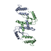
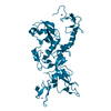
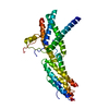
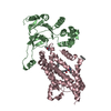
| Entry | Database: PDB / ID: 1e7d | ||||||
|---|---|---|---|---|---|---|---|
| Title | Endonuclease VII (ENDOVII) Ffrom Phage T4 | ||||||
 Components Components | RECOMBINATION ENDONUCLEASE VII | ||||||
 Keywords Keywords | HYDROLASE / ENDONUCLEASE / RESOLVASE / HOLLIDAY JUNCTION / DNASE | ||||||
| Function / homology |  Function and homology information Function and homology informationendonuclease activity / Hydrolases; Acting on ester bonds / hydrolase activity / metal ion binding Similarity search - Function | ||||||
| Biological species |  BACTERIOPHAGE T4 (virus) BACTERIOPHAGE T4 (virus) | ||||||
| Method |  X-RAY DIFFRACTION / MOLECULAR REPLACEMENT / Resolution: 2.8 Å X-RAY DIFFRACTION / MOLECULAR REPLACEMENT / Resolution: 2.8 Å | ||||||
 Authors Authors | Raaijmakers, H.C.A. / Vix, O. / Toro, I. / Suck, D. | ||||||
 Citation Citation | Journal: J.Mol.Biol. / Year: 2001 Title: Conformational Flexibility in T4 Endonuclease Vii Revealed by Crystallography: Implications for Substrate Binding and Cleavage Authors: Raaijmakers, H.C.A. / Toro, I. / Birkenbihl, R. / Kemper, B. / Suck, D. #1: Journal: Embo J. / Year: 1999Title: X-Ray Structure of T4 Endonuclease Vii: A DNA Junction Resolvase with a Novel Fold and Unusual Domain Swapped Dimer Architecture Authors: Raaijmakers, H. / Vix, O. / Toro, I. / Golz, S. / Kemper, B. / Suck, D. | ||||||
| History |
|
- Structure visualization
| Structure viewer | Molecule: MolmilJmol/JSmol |
|---|
- Downloads & links
-Download
| PDBx/mmCIF format | 1e7d.cif.gz | 77.2 KB | Display | PDBx/mmCIF format |
|---|---|---|---|---|
| PDB format | pdb1e7d.ent.gz | 57.4 KB | Display | PDB format |
| PDBx/mmJSON format | 1e7d.json.gz | Tree view | PDBx/mmJSON format | |
| Others |  Other downloads Other downloads |
-Validation report
| Arichive directory | https://data.pdbj.org/pub/pdb/validation_reports/e7/1e7dftp://data.pdbj.org/pub/pdb/validation_reports/e7/1e7d | HTTPS FTP |
|---|
-Related structure data
| Related structure data |  1e7lSC S: Starting model for refinement C: citing same article ( |
|---|---|
| Similar structure data |


-Links
 PDBj
PDBj- Assembly
| Deposited unit | 
| ||||||||
|---|---|---|---|---|---|---|---|---|---|
| 1 |
| ||||||||
| Unit cell |
| ||||||||
| Noncrystallographic symmetry (NCS) | NCS oper: (Code: given Matrix: (-0.4964, 0.8391, -0.2226), Vector: Details | BIOLOGICAL_UNIT: DIMERIC | |
-Components
| #1: Protein | Mass: 18173.777 Da / Num. of mol.: 2 Source method: isolated from a genetically manipulated source Details: ONE ZN BOUND TO CYS 23,26,58,61 OF EACH CHAIN CA LIGANDED TO ASP40 AND ASN 62 Source: (gene. exp.) BACTERIOPHAGE T4 (virus) / Cellular location: CYTOPLASM / Gene: GP49 / Plasmid: PET11A / Cellular location (production host): CYTOPLASM / Production host:  References: UniProt: P13340, crossover junction endodeoxyribonuclease #2: Chemical |   Mass: 65.409 Da / Num. of mol.: 2 / Source method: obtained synthetically / Formula: Zn Mass: 65.409 Da / Num. of mol.: 2 / Source method: obtained synthetically / Formula: Zn#3: Chemical |   Mass: 40.078 Da / Num. of mol.: 2 / Source method: obtained synthetically / Formula: Ca Mass: 40.078 Da / Num. of mol.: 2 / Source method: obtained synthetically / Formula: Ca#4: Chemical |   Mass: 35.453 Da / Num. of mol.: 3 / Source method: obtained synthetically / Formula: Cl Mass: 35.453 Da / Num. of mol.: 3 / Source method: obtained synthetically / Formula: Cl#5: Water | ChemComp-HOH / |  Mass: 18.015 Da / Num. of mol.: 30 / Source method: isolated from a natural source / Formula: H2O Mass: 18.015 Da / Num. of mol.: 30 / Source method: isolated from a natural source / Formula: H2O |
|---|
-Experimental details
-Experiment
| Experiment | Method: X-RAY DIFFRACTION / Number of used crystals: 1 |
|---|
- Sample preparation
| Crystal | Density Matthews: 2.18 Å3/Da / Density % sol: 44.16 % | ||||||||||||||||||||||||||||||||||||||||||||||||
|---|---|---|---|---|---|---|---|---|---|---|---|---|---|---|---|---|---|---|---|---|---|---|---|---|---|---|---|---|---|---|---|---|---|---|---|---|---|---|---|---|---|---|---|---|---|---|---|---|---|
| Crystal grow | Method: vapor diffusion, hanging drop / pH: 8.2 Details: HANGING DROP VAPOUR DIFFUSION DROP SIZE: 1 + 1 UL PROTEIN SOLUTION: 16 MG/ML ENDOVII, 175 MM NACL, 20 MM MGCL2, 2 MM ZNCL2, 10 MM 2-MERCAPTO-ETHANOL, 10 % GLYCEROL, 10 MM MOPS PH6.5. WELL: ...Details: HANGING DROP VAPOUR DIFFUSION DROP SIZE: 1 + 1 UL PROTEIN SOLUTION: 16 MG/ML ENDOVII, 175 MM NACL, 20 MM MGCL2, 2 MM ZNCL2, 10 MM 2-MERCAPTO-ETHANOL, 10 % GLYCEROL, 10 MM MOPS PH6.5. WELL: 16-18% PEG 5000 MME, 200 MM CACL2, 20 MM AMMONIUM SULPHATE, 10 MM 2-MERCAPTO-ETHANOL, 100 MM TRIS PH 8.2, C.A. 1 MM SODIUM AZIDE | ||||||||||||||||||||||||||||||||||||||||||||||||
| Crystal grow | *PLUS Temperature: 4 ℃ / pH: 8.2 / Method: vapor diffusion, hanging drop | ||||||||||||||||||||||||||||||||||||||||||||||||
| Components of the solutions | *PLUS
|
-Data collection
| Diffraction | Mean temperature: 100 K |
|---|---|
| Diffraction source | Source: ROTATING ANODE / Type: ENRAF-NONIUS / Wavelength: 1.5418 |
| Detector | Type: MARRESEARCH / Detector: IMAGE PLATE / Date: Jan 15, 1999 / Details: NI COATED MIRRORS |
| Radiation | Monochromator: NI COATED MIRRORS / Protocol: SINGLE WAVELENGTH / Monochromatic (M) / Laue (L): M / Scattering type: x-ray |
| Radiation wavelength | Wavelength: 1.5418 Å / Relative weight: 1 |
| Reflection | Resolution: 2.8→44.281 Å / Num. obs: 8966 / % possible obs: 99.8 % / Redundancy: 3.2 % / Rsym value: 0.096 / Net I/σ(I): 7.2 |
| Reflection shell | Resolution: 2.8→2.9 Å / Mean I/σ(I) obs: 3.2 / Rsym value: 0.283 / % possible all: 99.7 |
| Reflection | *PLUS Num. measured all: 28458 / Rmerge(I) obs: 0.096 |
| Reflection shell | *PLUS % possible obs: 99.7 % / Rmerge(I) obs: 0.283 |
- Processing
| Software |
| ||||||||||||||||||||||||||||||||||||||||||||||||||||||||||||||||||||||||||||||||||||
|---|---|---|---|---|---|---|---|---|---|---|---|---|---|---|---|---|---|---|---|---|---|---|---|---|---|---|---|---|---|---|---|---|---|---|---|---|---|---|---|---|---|---|---|---|---|---|---|---|---|---|---|---|---|---|---|---|---|---|---|---|---|---|---|---|---|---|---|---|---|---|---|---|---|---|---|---|---|---|---|---|---|---|---|---|---|
| Refinement | Method to determine structure: MOLECULAR REPLACEMENT Starting model: 1E7L Resolution: 2.8→10 Å / SU B: 7.34 / SU ML: 0.15 / Cross valid method: THROUGHOUT / σ(F): 0 / ESU R Free: 0.52 Details: TIGHT NCS RESTRAINTS SIMILAR TO ENTRY 1EN7, BUT IN A DIFFERENT SPACE GROUP THE ATOMS WITH ZERO OCCUPANCY ARE DISORDERED, AND THOSE WITH HALF OCCUPANCY ARE PARTLY DISORDERED
| ||||||||||||||||||||||||||||||||||||||||||||||||||||||||||||||||||||||||||||||||||||
| Refinement step | Cycle: LAST / Resolution: 2.8→10 Å
| ||||||||||||||||||||||||||||||||||||||||||||||||||||||||||||||||||||||||||||||||||||
| Refine LS restraints |
| ||||||||||||||||||||||||||||||||||||||||||||||||||||||||||||||||||||||||||||||||||||
| Software | *PLUS Name: REFMAC / Classification: refinement | ||||||||||||||||||||||||||||||||||||||||||||||||||||||||||||||||||||||||||||||||||||
| Refinement | *PLUS Lowest resolution: 27 Å / Num. reflection obs: 12364 / Rfactor obs: 0.245 | ||||||||||||||||||||||||||||||||||||||||||||||||||||||||||||||||||||||||||||||||||||
| Solvent computation | *PLUS | ||||||||||||||||||||||||||||||||||||||||||||||||||||||||||||||||||||||||||||||||||||
| Displacement parameters | *PLUS | ||||||||||||||||||||||||||||||||||||||||||||||||||||||||||||||||||||||||||||||||||||
| Refine LS restraints | *PLUS
|
