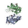
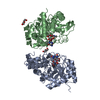
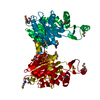
BIOMOLECULE: 1 THIS ENTRY CONTAINS THE CRYSTALLOGRAPHIC ASYMMETRIC UNIT WHICH CONSISTS OF 1 CHAINS. ... BIOMOLECULE: 1 THIS ENTRY CONTAINS THE CRYSTALLOGRAPHIC ASYMMETRIC UNIT WHICH CONSISTS OF 1 CHAINS. SEE REMARK 350 FOR INFORMATION ON GENERATING THE BIOLOGICAL MOLECULE(S). SIZE EXCLUSION CHROMATOGRAPHY WITH STATIC LIGHT SCATTERING SUPPORTS THE ASSIGNMENT OF A DIMER AS THE SIGNIFICANT OLIGOMERIZATION STATE.
Remark 999
SEQUENCE THE CONSTRUCT WAS EXPRESSED WITH A PURIFICATION TAG MGSDKIHHHHHHENLYFQG. THE TAG WAS ... SEQUENCE THE CONSTRUCT WAS EXPRESSED WITH A PURIFICATION TAG MGSDKIHHHHHHENLYFQG. THE TAG WAS REMOVED WITH TEV PROTEASE LEAVING ONLY A GLYCINE (0) FOLLOWED BY THE TARGET SEQUENCE.
Resolution: 2→27.587 Å / Num. obs: 23426 / % possible obs: 91.2 % / Biso Wilson estimate: 33.272 Å2 / Rmerge(I) obs: 0.063 / Net I/σ(I): 7.47
Reflection shell
Diffraction-ID: 1
Resolution (Å)
Highest resolution (Å)
Rmerge(I) obs
Mean I/σ(I) obs
Num. measured obs
Num. unique all
% possible all
2-2.07
0.351
2.6
3345
3879
83.5
2.07-2.15
0.3
2.6
3710
4036
89.1
2.15-2.25
0.247
3.4
3696
4179
87.2
2.25-2.37
0.196
3.7
4070
4364
91.5
2.37-2.52
0.157
4.4
4263
4454
93
2.52-2.71
0.123
5.5
3991
4203
92.3
2.71-2.99
0.088
7.2
4423
4555
94.6
2.99-3.42
0.062
10.1
4318
4415
94.1
3.42
0.038
15.7
4204
4323
92.7
-
Phasing
Phasing
Method: MAD
-
Processing
Software
Name
Version
Classification
NB
MolProbity
3beta29
modelbuilding
SHELX
phasing
REFMAC
5.2.0019
refinement
XSCALE
datascaling
PDB_EXTRACT
2
dataextraction
MAR345
CCD
datacollection
XDS
datareduction
SHELXD
phasing
autoSHARP
phasing
Refinement
Method to determine structure: MAD / Resolution: 2→27.587 Å / Cor.coef. Fo:Fc: 0.952 / Cor.coef. Fo:Fc free: 0.919 / SU B: 10.253 / SU ML: 0.138 / TLS residual ADP flag: LIKELY RESIDUAL / Cross valid method: THROUGHOUT / σ(F): 0 / ESU R: 0.184 / ESU R Free: 0.167 Stereochemistry target values: MAXIMUM LIKELIHOOD WITH PHASES Details: 1. HYDROGENS HAVE BEEN ADDED IN THE RIDING POSITIONS. 2. A MET-INHIBITION PROTOCOL WAS USED FOR SELENOMETHIONINE INCORPORATION DURING PROTEIN EXPRESSION. THE OCCUPANCY OF THE SE ATOMS IN THE ...Details: 1. HYDROGENS HAVE BEEN ADDED IN THE RIDING POSITIONS. 2. A MET-INHIBITION PROTOCOL WAS USED FOR SELENOMETHIONINE INCORPORATION DURING PROTEIN EXPRESSION. THE OCCUPANCY OF THE SE ATOMS IN THE MSE RESIDUES WAS REDUCED TO 0.75 FOR THE REDUCED SCATTERING POWER DUE TO PARTIAL S-MET INCORPORATION. 3. RESIDUES 159-181 ARE DISORDERED AND ARE NOT INCLUDED IN THE MODEL. 4. A GLYCEROL MOLECULE FROM THE CRYO SOLUTION IS MODELED. 5. ATOM RECORDS CONTAIN RESIDUAL B FACTORS ONLY.
Rfactor
Num. reflection
% reflection
Selection details
Rfree
0.239
1200
5.1 %
RANDOM
Rwork
0.192
-
-
-
all
0.195
-
-
-
obs
0.195
23425
97.7 %
-
Solvent computation
Ion probe radii: 0.8 Å / Shrinkage radii: 0.8 Å / VDW probe radii: 1.2 Å / Solvent model: BABINET MODEL WITH MASK
Displacement parameters
Biso mean: 28.712 Å2
Baniso -1
Baniso -2
Baniso -3
1-
0.49 Å2
0 Å2
-0.84 Å2
2-
-
-0.55 Å2
0 Å2
3-
-
-
-0.87 Å2
Refinement step
Cycle: LAST / Resolution: 2→27.587 Å
Protein
Nucleic acid
Ligand
Solvent
Total
Num. atoms
2390
0
6
165
2561
Refine LS restraints
Refine-ID
Type
Dev ideal
Dev ideal target
Number
X-RAY DIFFRACTION
r_bond_refined_d
0.013
0.022
2468
X-RAY DIFFRACTION
r_bond_other_d
0.003
0.02
1632
X-RAY DIFFRACTION
r_angle_refined_deg
1.622
1.981
3357
X-RAY DIFFRACTION
r_angle_other_deg
1.306
3
4003
X-RAY DIFFRACTION
r_dihedral_angle_1_deg
3.951
5
316
X-RAY DIFFRACTION
r_dihedral_angle_2_deg
34.793
24.151
106
X-RAY DIFFRACTION
r_dihedral_angle_3_deg
10.878
15
417
X-RAY DIFFRACTION
r_dihedral_angle_4_deg
10.831
15
13
X-RAY DIFFRACTION
r_chiral_restr
0.091
0.2
392
X-RAY DIFFRACTION
r_gen_planes_refined
0.006
0.02
2746
X-RAY DIFFRACTION
r_gen_planes_other
0.002
0.02
491
X-RAY DIFFRACTION
r_nbd_refined
0.168
0.2
439
X-RAY DIFFRACTION
r_nbd_other
0.14
0.2
1608
X-RAY DIFFRACTION
r_nbtor_refined
0.152
0.2
1190
X-RAY DIFFRACTION
r_nbtor_other
0.072
0.2
1266
X-RAY DIFFRACTION
r_xyhbond_nbd_refined
0.099
0.2
121
X-RAY DIFFRACTION
r_symmetry_vdw_refined
0.121
0.2
15
X-RAY DIFFRACTION
r_symmetry_vdw_other
0.162
0.2
41
X-RAY DIFFRACTION
r_symmetry_hbond_refined
0.086
0.2
3
X-RAY DIFFRACTION
r_mcbond_it
1.562
3
1594
X-RAY DIFFRACTION
r_mcbond_other
0.368
3
630
X-RAY DIFFRACTION
r_mcangle_it
2.351
5
2494
X-RAY DIFFRACTION
r_scbond_it
4.548
8
993
X-RAY DIFFRACTION
r_scangle_it
6.094
11
858
LS refinement shell
Resolution: 2→2.052 Å / Total num. of bins used: 20
Rfactor
Num. reflection
% reflection
Rfree
0.245
83
-
Rwork
0.271
1652
-
obs
-
1735
97.91 %
Refinement TLS params.
Method: refined / Origin x: -0.365 Å / Origin y: 39.03 Å / Origin z: 46.118 Å
11
12
13
21
22
23
31
32
33
T
-0.0665 Å2
0.0007 Å2
0.0033 Å2
-
-0.0425 Å2
-0.0013 Å2
-
-
-0.034 Å2
L
0.8101 °2
-0.1356 °2
-0.0175 °2
-
0.5288 °2
-0.0538 °2
-
-
1.111 °2
S
-0.0013 Å °
-0.0117 Å °
0.0877 Å °
-0.0241 Å °
0.0375 Å °
-0.0051 Å °
-0.0866 Å °
-0.0575 Å °
-0.0361 Å °
Refinement TLS group
Refine-ID: X-RAY DIFFRACTION / Refine TLS-ID: 1 / Selection: ALL / Auth asym-ID: A / Label asym-ID: A
ID
Auth seq-ID
Label seq-ID
1
0 - 158
1 - 159
2
182 - 330
183 - 331
+
About Yorodumi
-
News
-
Feb 9, 2022. New format data for meta-information of EMDB entries
New format data for meta-information of EMDB entries
Version 3 of the EMDB header file is now the official format.
The previous official version 1.9 will be removed from the archive.
In the structure databanks used in Yorodumi, some data are registered as the other names, "COVID-19 virus" and "2019-nCoV". Here are the details of the virus and the list of structure data.
Jan 31, 2019. EMDB accession codes are about to change! (news from PDBe EMDB page)
EMDB accession codes are about to change! (news from PDBe EMDB page)
The allocation of 4 digits for EMDB accession codes will soon come to an end. Whilst these codes will remain in use, new EMDB accession codes will include an additional digit and will expand incrementally as the available range of codes is exhausted. The current 4-digit format prefixed with “EMD-” (i.e. EMD-XXXX) will advance to a 5-digit format (i.e. EMD-XXXXX), and so on. It is currently estimated that the 4-digit codes will be depleted around Spring 2019, at which point the 5-digit format will come into force.
The EM Navigator/Yorodumi systems omit the EMD- prefix.
Related info.:Q: What is EMD? / ID/Accession-code notation in Yorodumi/EM Navigator
Yorodumi is a browser for structure data from EMDB, PDB, SASBDB, etc.
This page is also the successor to EM Navigator detail page, and also detail information page/front-end page for Omokage search.
The word "yorodu" (or yorozu) is an old Japanese word meaning "ten thousand". "mi" (miru) is to see.
Related info.:EMDB / PDB / SASBDB / Comparison of 3 databanks / Yorodumi Search / Aug 31, 2016. New EM Navigator & Yorodumi / Yorodumi Papers / Jmol/JSmol / Function and homology information / Changes in new EM Navigator and Yorodumi
 Movie
Movie Controller

 Yorodumi
 Open data
 Basic information
 Components
 Keywords
 Function and homology information
 Exiguobacterium sibiricum (bacteria)
 X-RAY DIFFRACTION /
  Authors
 Citation
 Structure visualization
 Downloads & links
 Other downloads
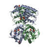


 PDBj
PDBj


 Assembly

 Mass: 92.094 Da / Num. of mol.: 1 / Source method: obtained synthetically / Formula: C3H8O3
 Mass: 18.015 Da / Num. of mol.: 165 / Source method: isolated from a natural source / Formula: H2O
 Sample preparation
 / Beamline: BL9-2 / Wavelength: 0.91837, 0.97920
 Processing