- PDB-3c9h: Crystal structure of the substrate binding protein of the ABC tra... -
+
Open data
ID or keywords:
Loading...
-
Basic information
Entry
Database: PDB / ID: 3c9h
Title
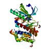
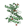
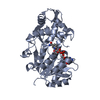
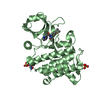
Crystal structure of the substrate binding protein of the ABC transporter from Agrobacterium tumefaciens
Components
ABC transporter, substrate binding protein
Keywords
TRANSPORT PROTEIN / substrate binding protein / ABC transporter / Structural genomics / MCSG / PSI-2 / Protein Structure Initiative / Midwest Center for Structural Genomics
Monochromator: Si(111) channel / Protocol: SINGLE WAVELENGTH / Monochromatic (M) / Laue (L): M / Scattering type: x-ray
Radiation wavelength
Wavelength: 0.9794 Å / Relative weight: 1
Reflection
Resolution: 1.9→68.2 Å / Num. all: 54496 / Num. obs: 54136 / % possible obs: 99.34 % / Observed criterion σ(I): 2 / Redundancy: 8.7 % / Biso Wilson estimate: 25 Å2 / Rmerge(I) obs: 0.112 / Net I/σ(I): 19.78
Reflection shell
Resolution: 1.9→1.95 Å / Redundancy: 8.2 % / Rmerge(I) obs: 0.731 / Mean I/σ(I) obs: 1.57 / Num. unique all: 4178 / % possible all: 97.77
-
Processing
Software
Name
Version
Classification
REFMAC
5.2.0019
refinement
SBC-Collect
datacollection
HKL-3000
datareduction
HKL-3000
datascaling
MLPHARE
phasing
Refinement
Method to determine structure: SAD / Resolution: 1.9→35.6 Å / Cor.coef. Fo:Fc: 0.962 / Cor.coef. Fo:Fc free: 0.937 / SU B: 7.333 / SU ML: 0.098 / Cross valid method: THROUGHOUT / σ(F): 0 / σ(I): 0 / ESU R: 0.497 / ESU R Free: 0.144 Stereochemistry target values: MAXIMUM LIKELIHOOD WITH PHASES Details: HYDROGENS HAVE BEEN ADDED IN THE RIDING POSITIONS
Rfactor
Num. reflection
% reflection
Selection details
Rfree
0.22817
2898
5.1 %
RANDOM
Rwork
0.17259
-
-
-
all
0.17539
54136
-
-
obs
0.17539
54136
99.34 %
-
Solvent computation
Ion probe radii: 0.8 Å / Shrinkage radii: 0.8 Å / VDW probe radii: 1.2 Å / Solvent model: MASK
Displacement parameters
Biso mean: 24.183 Å2
Baniso -1
Baniso -2
Baniso -3
1-
-0.62 Å2
0 Å2
0 Å2
2-
-
1.49 Å2
0 Å2
3-
-
-
-0.87 Å2
Refinement step
Cycle: LAST / Resolution: 1.9→35.6 Å
Protein
Nucleic acid
Ligand
Solvent
Total
Num. atoms
5265
0
28
433
5726
Refine LS restraints
Refine-ID
Type
Dev ideal
Dev ideal target
Number
X-RAY DIFFRACTION
r_bond_refined_d
0.013
0.022
5415
X-RAY DIFFRACTION
r_bond_other_d
0.001
0.02
3672
X-RAY DIFFRACTION
r_angle_refined_deg
1.371
1.956
7356
X-RAY DIFFRACTION
r_angle_other_deg
0.934
3
8897
X-RAY DIFFRACTION
r_dihedral_angle_1_deg
5.94
5
669
X-RAY DIFFRACTION
r_dihedral_angle_2_deg
34.946
23.306
245
X-RAY DIFFRACTION
r_dihedral_angle_3_deg
13.921
15
871
X-RAY DIFFRACTION
r_dihedral_angle_4_deg
15.977
15
42
X-RAY DIFFRACTION
r_chiral_restr
0.084
0.2
802
X-RAY DIFFRACTION
r_gen_planes_refined
0.005
0.02
6058
X-RAY DIFFRACTION
r_gen_planes_other
0.001
0.02
1136
X-RAY DIFFRACTION
r_nbd_refined
0.218
0.2
1185
X-RAY DIFFRACTION
r_nbd_other
0.198
0.2
3819
X-RAY DIFFRACTION
r_nbtor_refined
0.179
0.2
2594
X-RAY DIFFRACTION
r_nbtor_other
0.086
0.2
2743
X-RAY DIFFRACTION
r_xyhbond_nbd_refined
0.176
0.2
376
X-RAY DIFFRACTION
r_xyhbond_nbd_other
0.016
0.2
1
X-RAY DIFFRACTION
r_symmetry_vdw_refined
0.167
0.2
9
X-RAY DIFFRACTION
r_symmetry_vdw_other
0.277
0.2
51
X-RAY DIFFRACTION
r_symmetry_hbond_refined
0.133
0.2
19
X-RAY DIFFRACTION
r_mcbond_it
1.486
1.5
4314
X-RAY DIFFRACTION
r_mcbond_other
0.544
1.5
1351
X-RAY DIFFRACTION
r_mcangle_it
1.726
2
5399
X-RAY DIFFRACTION
r_scbond_it
3.033
3
2439
X-RAY DIFFRACTION
r_scangle_it
4.11
4.5
1957
LS refinement shell
Resolution: 1.9→1.949 Å / Total num. of bins used: 20
Rfactor
Num. reflection
% reflection
Rfree
0.338
206
-
Rwork
0.235
3879
-
obs
-
4085
97.77 %
Refinement TLS params.
Method: refined / Origin x: 37.753 Å / Origin y: -0.084 Å / Origin z: 11.145 Å
11
12
13
21
22
23
31
32
33
T
-0.0293 Å2
0.0063 Å2
-0.0003 Å2
-
-0.0228 Å2
0.0024 Å2
-
-
-0.0462 Å2
L
0.6245 °2
0.1767 °2
-0.0115 °2
-
0.1016 °2
-0.0221 °2
-
-
0.2935 °2
S
0.0131 Å °
0.0196 Å °
0.0044 Å °
-0.0151 Å °
0.0184 Å °
-0.0024 Å °
-0.0121 Å °
-0.0162 Å °
-0.0314 Å °
Refinement TLS group
ID
Refine-ID
Refine TLS-ID
Auth asym-ID
Label asym-ID
Auth seq-ID
Label seq-ID
1
X-RAY DIFFRACTION
1
A
A
16 - 60
16 - 60
2
X-RAY DIFFRACTION
1
A
A
61 - 100
61 - 100
3
X-RAY DIFFRACTION
1
A
A
101 - 150
101 - 150
4
X-RAY DIFFRACTION
1
A
A
151 - 200
151 - 200
5
X-RAY DIFFRACTION
1
A
A
201 - 250
201 - 250
6
X-RAY DIFFRACTION
1
A
A
251 - 300
251 - 300
7
X-RAY DIFFRACTION
1
A
A
301 - 354
301 - 354
8
X-RAY DIFFRACTION
1
B
B
15 - 60
15 - 60
9
X-RAY DIFFRACTION
1
B
B
61 - 100
61 - 100
10
X-RAY DIFFRACTION
1
B
B
101 - 150
101 - 150
11
X-RAY DIFFRACTION
1
B
B
151 - 200
151 - 200
12
X-RAY DIFFRACTION
1
B
B
201 - 250
201 - 250
13
X-RAY DIFFRACTION
1
B
B
251 - 300
251 - 300
14
X-RAY DIFFRACTION
1
B
B
301 - 353
301 - 353
+
About Yorodumi
-
News
-
Feb 9, 2022. New format data for meta-information of EMDB entries
New format data for meta-information of EMDB entries
Version 3 of the EMDB header file is now the official format.
The previous official version 1.9 will be removed from the archive.
In the structure databanks used in Yorodumi, some data are registered as the other names, "COVID-19 virus" and "2019-nCoV". Here are the details of the virus and the list of structure data.
Jan 31, 2019. EMDB accession codes are about to change! (news from PDBe EMDB page)
EMDB accession codes are about to change! (news from PDBe EMDB page)
The allocation of 4 digits for EMDB accession codes will soon come to an end. Whilst these codes will remain in use, new EMDB accession codes will include an additional digit and will expand incrementally as the available range of codes is exhausted. The current 4-digit format prefixed with “EMD-” (i.e. EMD-XXXX) will advance to a 5-digit format (i.e. EMD-XXXXX), and so on. It is currently estimated that the 4-digit codes will be depleted around Spring 2019, at which point the 5-digit format will come into force.
The EM Navigator/Yorodumi systems omit the EMD- prefix.
Related info.:Q: What is EMD? / ID/Accession-code notation in Yorodumi/EM Navigator
Yorodumi is a browser for structure data from EMDB, PDB, SASBDB, etc.
This page is also the successor to EM Navigator detail page, and also detail information page/front-end page for Omokage search.
The word "yorodu" (or yorozu) is an old Japanese word meaning "ten thousand". "mi" (miru) is to see.
Related info.:EMDB / PDB / SASBDB / Comparison of 3 databanks / Yorodumi Search / Aug 31, 2016. New EM Navigator & Yorodumi / Yorodumi Papers / Jmol/JSmol / Function and homology information / Changes in new EM Navigator and Yorodumi
 Movie
Movie Controller

 Yorodumi
 Open data
 Basic information
 Components
 Keywords
 Function and homology information
 Agrobacterium tumefaciens str. (bacteria)
 X-RAY DIFFRACTION /
  Authors
 Citation
 Structure visualization
 Downloads & links
 Other downloads


 PDBj
PDBj


 Assembly


 Mass: 24.305 Da / Num. of mol.: 2 / Source method: obtained synthetically / Formula: Mg
 Mass: 192.124 Da / Num. of mol.: 2 / Source method: obtained synthetically / Formula: C6H8O7
 Mass: 18.015 Da / Num. of mol.: 433 / Source method: isolated from a natural source / Formula: H2O
 Sample preparation
 / Beamline: 19-ID / Wavelength: 0.9794 Å
 Processing